What Do Viruses Like HIV & Corona Have In Common With Exosomes?
Updated: Jun 5, 2020
Authors: Robert O. Young CPC, MSc, DSc, PhD, Naturopathic Practitioner
Galina Migalko MD, NMD
Dr. James Hildreth PhD MD, proposed that “the virus is fully an exosome in every sense of the word.” [1]

Abstract
There is only one sickness, one disease and one treatment. The one sickness and one disease is the over-acidification of the blood and then interstitial fluids due to an inverted way of living, eating, drinking, breathing, thinking, feeling and believing. There are six major contributing factors that lead to the declining acidic pH of the body fluids. As the pH of the body fluids become compensated by these six contributing factors and the body cell membranes and genetic material begin to degenerate the cells release exosomes as a defense to activate and support the lymphocytes to release oxygen species or antioxidants to reduce the acidic loads stored in the interstitial fluids of the Interstitium. The one treatment is to support the immune system with increased amounts of reduced oxygen (O-) and reduced hydrogen (H-) to restore the alkaline design of the body fluids, open up the channels of elimination in order to remove dietary, metabolic, respiratory and environmental toxic acidic waste held in the interstial fluids of the Interstitium and thus restoring health, energy and vitality to the body.

Introduction
What Are Exosomes?

Exosomes are membrane bound extracellular vesicles (EVs) that are produced in the endosomal compartment of most eukaryotic cells.[2][3][4] The multivesicular body (MVB) is an endosome defined by intraluminal vesicles (ILVs) that bud inward into the endosomal lumen. If the MVB fuses with the cell surface (the plasma membrane), these ILVs are released as exosomes. In multicellular organisms, exosomes and other EVs are present in cells that make up tissues and can also be found in biological fluids including intracellular fluids, intravascular fluids, interstitial fluids, urine, and cerebrospinal fluid. They are also released in vitro by cultured cells into their growth medium.[5][6][7][8]
Since the size of exosomes is limited by that of the parent MVB, exosomes are generally thought to be smaller than most other EVs, from about 30 to 150 nanometres (nm) in diameter: around the same size as many lipoproteins but much smaller than cells.[5] Compared with EVs in general, it is becoming more clear that exosomes do have unique characteristics or functions and can be separated or distinguished effectively from other EVs.[2] EVs including exosomes carry markers of cells of origin and have specialized functions in physiological processes, from coagulation and intercellular signaling to acidic waste management of the intravascular and interstitial fluids of the Interstitium – the largest organ of the human body.[5]
Are Exosomes Viruses?
There is NO scientific evidence from ANY research (published or otherwise) from ANY scientist or group of scientists any where in the World validating the existence of the so-called invisible so-called virus or that exosomes have been proven to be the virus.[29]
Exosomes are created endogenously by the cells, even the red blood cells as a means of mediating or buffering metabolic, environmental, dietary and/or respiratory acidic waste in order to maintain the delicate pH balance of the intravascular fluids, the interstitial fluids and the intracellular fluids of the body cells at 7.365.[9][31]
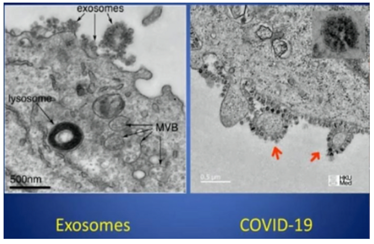
Exosomes and the so-called Corona virus or COVOD-2 and 19 also referred to as the SARS virus are identical in appearance and size and have the same ACE-2 receptor sites, containing the same RNA found in the interstitial fluids of the Interstitium surrounding the cells of the bronchoalveolar. The exosome or the so-called Corona virus is created endogenously and is NOT transmittable or contagious unless transmitted by injection from the isolated exosome(s) of one person or animal to another person.

Are COVID-19 and HIV Exosomes?
Based upon electron microscopy the so-called COVID-19 virus and the the so-called HIV virus are 100 nm in diameter and appear identical in structure to the exosome.


On January 18th, of 2020, three scientists published a scientific paper describing the protective purpose of exosomes, entitled, “Exosome-Mediated Transfer of ACE2 (Angiotensin-Converting Enzyme 2) from Endothelial Progenitor Cells Promotes Survival and Function of Endothelial Cell.”[9]

Research on Exosomes and Their Support of the Lymphocytes (Immune System) in Reducing Cancer-causing Acidic Waste
Exosomes from red blood cells contain the transferrin receptor which is absent in mature erythrocytes. Dendritic cell-derived exosomes express MHC I, MHC II, and costimulatory molecules and have been proven to be able to induce and enhance antigen-specific T-cell responses in vivo in reducing metabolic, dietary, environmental and respiratory acidic waste deposited into the interstitial fluids of the Interstitium.[10]

What Is the Relationship Between Exosomes and COVID-19
They both contain the ACE2, or angiotensin converting enzyme-2 receptor and visually, using an electron microscope measure the same size. The exosomes or should we say the COVID-19, ACE2 receptor chops up two forms of a protein called angiotensin to keep blood pressure stable by protecting cell membranes from cellular breakdown from metabolic, dietary, environmental and respiratory acidic waste.[9]
So What is Causing the Symptoms of COVID-19 and the Release of Exosomes into the Extracellular Fluid Matrix?
It has come down to a four letter word – ACID! So where is the ACID or toxics coming from?
The seven major contributing factors that cause cellular breakdown and the release of exosomes into the extracellular matrix are as follows:

1. Electro-magnetic pulsating frequencies ranging from 1GHz to 600GHz.[11][12][13]

2. Carbon Dioxide and Monoxide Poisoning.[14][15][16][17][18][19]
3. Pathological blood coagulation leading to hypoxia, interstitial fluid lung disease, and then sepsis.[20]
4. Glyphosate Acid Poisoning from non-organic fruit and vegetables.[21][22]
5. Lactic Acid Poisoning from diet and metabolism.[21]22]
6. Uric, Nitric, Sulphuric and Phosphoric Acid Poisoning from eating the flesh and blood of animals.[21][22]
7. Genetically Modified Organisms in our food supply and vaccines.[21][22] [Please see the comparisons of flu vaccine frequency in older people with Covid-19 mortality, using figures fromVaccines Today EU and Worldometer (13 May 2020):[23]

Among countries omitted in the EU data was Belgium, which has the highest Covid-19 mortality rate in the world, at 763 per million. While specific data for older people were not available on the official Belgian statistics website, flu vaccination coverage of the overall population is relatively high.

Globally the highest uptake of the flu vaccine by seniors in 2018-2019 was in South Korea, at 83%. Third (after the UK) was the USA with 68%, and fourth was New Zealand with 67%.
Vaccines may be a contributing factor to other so-called viral conditions . As reported in , whooping cough outbreaks have infected vaccinated as well as unvaccinated people.[24][25] Mandating of the chickenpox vaccine in the USA appears to have weakened the immunity gained from the naturally-acquired disease; a review by Goldman and King inVaccine journal showed increasing incidence of shingles.[26] Studies have indicated that people receiving the flu vaccine in one year were more likely to contract the H1N1 strain of exosomes in the following year.[27][28]

Despite some contrary cases, it is significant that the countries with highest death rates (Belgium, Spain, Italy, UK, France, Netherlands, Sweden, Ireland and USA) had all vaccinated at least half of their elderly population against flu. Denmark and Germany, with lower use of the flu vaccine, have considerably lower Covid-19 mortality. These patterns override interventions to curtail Covid-19: Sweden and Ireland have similar mortality but the former remained open for business while the other imposed strict lockdown.
How Can I Support the Alkaline Design of the Body and Reduce Metabolic, Dietary, Environmental and Respiratory Acidic Waste that is Making Me Sick and Tired?
First, read seven books by Dr. Robert O. Young to starting with Sick and Tired, Reclaim Your Inner Terrain –

1. Sick and Tired, Reclaim Your Inner Terrain[20]
\
2. The pH Miracle revised and updated[21]

3. Chlorine Dioxide (CLO2) As a Non-Toxic Antimicrobial Agent for Virus, Bacteria and Yeast (Candids Albicans)[29]

4. Alkalizing Nutritional Therapy in the Prevention and Treatment of any Cancerous Condition,[30]

5. Second Thoughts about Viruses, Vaccines, and the HIV/AIDS Hypothesis.[31]

6. The Possible Cause of Polio – Post-Polio, CNS, PViPD, Legionnaires, AIDS, and the Cancer Epidemic – Mass Acidic Chemical Poisoning?[32] and,

7. Pathological Blood Coagulation, The Mycotoxic Stress Test.[33]

Second, follow the protocol as outlined in Chapter 5 and 11 in The pH Miracle Revised and Updated for at least 12 weeks.[21]
Third, If you need further clarification and support you can setup a consultation with Dr. Robert O. Young by clicking here: https://www.drrobertyoung.com/services-page

Fourth, you can attend a pH Miracle Retreat and immerse yourself in a paradise of alkalinity. To learn more go to: www.phmiracleretreat.com

Methodology
In order to obtain the micrographs of exosomes or so-called viruses RT electronmicroscopy was used in addition to phase and dark field microscopy on the intravascular and interstitial fluids of the extracellular fluid matrix. We also used a unique patent-pending electron device for measuring the biochemistry, including the pH for analytical comparisons of the intracellular fluid matrix which include the intravascular and interstitial fluids of the Interstitium, the largest organ of the human body. By measuring all the body fluids for their biochemistry it was clear that patients who tested positive for HIV and Cornavirus where in decompensated acidosis of the interstitial fluids, including the interstitial fluids of the lungs.[20][31]
Conclusion
Exosomes are the so-called viruses since viruses have never been identified, isolated, purified, and cultured based upon the scientific method called Koch’s Postulates.[34] Exosomes are released endogenously from the body cells to assist in activating the immune response due to decompensated acidosis of the interstitial fluids of the Interstitium. Decompensated acidosis of the body fluids causes cell membrane degeneration and genetic mutation leading to all sicknesses and diseases. There are at least seven major contributing toxic factors that cause the increased levels of acidity in the body fluids leading to a significant decline in the alkaline design of the major body fluids (interstitial fluids of the Interstitium organ and the intravascular fludis) from their ideal pH of 7.365 to an unhealthy pH of 7.265 to 7.165. The seven major contributing acidic factors that cause all sickness and disease are:
1) pulsating electro-magnetic fields from satellites, cell phones, computers, cell towers, WiFi, electric cars, TV’s, etc.,
2) carbon dioxide and carbon monoxide poisoning from air-pollution,
3) pathological blood coagulation leading to hypoxia, interstitial lung disease and sepsis,
4) glyphosate poisoning from food, water and vaccines,
5) lactic acid poisoning from diet and metabolism,
6) uric, nitric, sulphuric and phosphoric acid poisoning from indigestion of eggs, fish, beef, chicken and pork, and finally,
7) the introduction of genetically modified organisms and aluminum oxide poisoning from vaccines and chem trails.[27-31]
References
[2] Théry C, Witwer KW, Aikawa E, Alcaraz MJ, Anderson JD, Andriantsitohaina R, et al. (2018). “Minimal information for studies of extracellular vesicles 2018 (MISEV2018): a position statement of the International Society for Extracellular Vesicles and update of the MISEV2014 guidelines”. Journal of Extracellular Vesicles. 7 (1): 1535750. doi:10.1080/20013078.2018.1535750. PMC 6322352. PMID 30637094.
[3] Yáñez-Mó M, Siljander PR, Andreu Z, Zavec AB, Borràs FE, Buzas EI, Buzas K, et al. (2015). “Biological properties of extracellular vesicles and their physiological functions”. Journal of Extracellular Vesicles. 4: 27066. doi:10.3402/jev.v4.27066. PMC 4433489. PMID 25979354.
[4] van Niel G, D’Angelo G, Raposo G (April 2018). “Shedding light on the cell biology of extracellular vesicles”. Nature Reviews. Molecular Cell Biology. 19 (4): 213–228. doi:10.1038/nrm.2017.125. PMID 29339798.
[5] van der Pol E, Böing AN, Harrison P, Sturk A, Nieuwland R (July 2012). “Classification, functions, and clinical relevance of extracellular vesicles”. Pharmacological Reviews. 64 (3): 676–705. doi:10.1124/pr.112.005983. PMID 22722893.
[6] Keller S, Sanderson MP, Stoeck A, Altevogt P (November 2006). “Exosomes: from biogenesis and secretion to biological function”. Immunology Letters. 107 (2): 102–8. doi:10.1016/j.imlet.2006.09.005. PMID 17067686.
[7] Spaull R, McPherson B, Gialeli A, Clayton A, Uney J, Heep A, Cordero-Llana Ó (April 2019). “Exosomes populate the cerebrospinal fluid of preterm infants with post-haemorrhagic hydrocephalus”. International Journal of Developmental Neuroscience. 73: 59–65. doi:10.1016/j.ijdevneu.2019.01.004. PMID 30639393.
[8] Dhondt B, Van Deun J, Vermaerke S, de Marco A, Lumen N, De Wever O, Hendrix A (June 2018). “Urinary extracellular vesicle biomarkers in urological cancers: From discovery towards clinical implementation”. The International Journal of Biochemistry & Cell Biology. 99: 236–256. doi:10.1016/j.biocel.2018.04.009. PMID 29654900.
[9] Wang J, Chen S, Bihl J, “Exosome-Mediated Transfer of ACE2 (Angiotensin-Converting Enzyme 2) from Endothelial Progenitor Cells Promotes Survival and Function.” Oxid Med Cell Longev, 2020 Jan 18;2020:4213541. doi: 10.1155/2020/4213541
[10] Mignot G, Roux S, Thery C, Ségura E, Zitvogel L (2006). “Prospects for exosomes in immunotherapy of cancer”. Journal of Cellular and Molecular Medicine. 10 (2): 376–88. doi:10.1111/j.1582-4934.2006.tb00406.x. PMC 3933128. PMID 16796806.
[11] Rubik, B. Bioelectromagnetic Medicine. Administrative Radiology Journal XVI(8), August 1997, 38-46.
[12] Young, R.O., “The Effects of ElectroMagnetic Frequencies (EMF) on the Blood and Biological Terrain.” https://www.drrobertyoung.com/…/the-effects-electromagnet-f…
[13] Young, R.O., “Adverse Health Effects of 5G Mobile Networking Technology Under Real-Life Conditions.” April 19th, 2020. https://www.drrobertyoung.com/…/adverse-health-effects-of-5…
[14] NOAA. (2016). In a high carbon dioxide world, dangerous waters ahead. (accessed on August 6, 2019)
[15] NOAA. (2018). What is Ocean Acidification? (accessed on August 6, 2019)
[16] National Geographic. (2017). Ocean Acidification. (accessed on August 6, 2019)
[17] NOAA. (2010). Ocean Acidification, Today and in the Future. (accessed on August 6, 2019)
[18] Young, R.O., Young, S.R, “The pH Miracle Revised and Updated.” Hachett Publishing, 2010.
[19] Young, R.O., Are the Interstitial Fluids Raining Acid on YOUR Lung Cells? (December 17th, 2019)
[20] Young, R.O., Migalko, G., “Interstitial Fluid Lung Disease (IFLD) of the Interstitium Organ the Cause and Self-Care to a Self-Cure for Lung Disease”. International Journal of Cancer Research & Therapy, https://bit.ly/2xD8VBP, January 20, 2020.
[21] Young, R.O., “Sick and Tired.” https://www.phmiracleproducts.com/…/books-audio-video/produ…
[22] Young, R.O., Young, S.R. “The pH Miracle Revised and Updated.” Grand Central Publishing, NY, NY, 2010. https://www.phmiracleproducts.com/…/the-ph-miracle-revised-
[23] Vaccines Today, EUandWorldometer (13 May 2020).
[24] Althouse, B.M., Scarpino, S.V. Asymptomatic transmission and the resurgence ofBordetella pertussis.BMC Med 13,146 (2015). https://doi.org/10.1186/s12916-015-0382-8…
[25] Zhang Q, Yin Z, Li Y, Luo H, Shao Z, Gao Y, et al.Prevalence of asymptomatic Bordetella pertussis and Bordetella parapertussis infections among school children in China as determined by pooled real-time PCR: A cross-sectional study. Scand J Infect Dis. 2014; 46:280–7.
[26] Goldman GS, King PG, “Review of the United States universal varicella vaccination program: Herpes zoster incidence rates, cost-effectiveness, and vaccine efficacy based primarily on the Antelope Valley Varicella Active Surveillance Project data.” Vaccine, 2013 Mar 25;31(13):1680-94. doi: 10.1016/j.vaccine.2012.05.050. Epub 2012 Jun 1.
[27] Cécile Viboud, Lone Simonsen, “Does Seasonal Influenza Vaccination Increase the Risk of Illness with the 2009 A/H1N1 Pandemic Virus?” PLoS Med. 2010 Apr; 7(4): e1000259.
[28] Belongia EA, Skowronski DM, McLean HQ, Chambers C, Sundaram ME, De Serres G, “Repeated annual influenza vaccination and vaccine effectiveness: review of evidence.” Expert Rev Vaccines. 2017 Jul;16(7):1-14. doi: 10.1080/14760584.2017.1334554. Epub 2017 Jun 9.
[29] Young, R.O.,”Chlorine Dioxide (CLO2) As a Non-Toxic Antimicrobial Agent for Virus, Bacteria and Yeast (Candids Albicans),” Hikari Omni Media, August 2nd, 2016. https://www.phmiracleproducts.com/…/chlorine-dioxide-clo2-b…
[30] Young, R.O., Migalko, G., “Alkalizing Nutritional Therapy in the Prevention and Treatment of any Cancerous Condition.” Hikari Omni Media, August 1st, 2016. https://www.phmiracleproducts.com/…/alkalizing-nutritional-…
[31] Young, R.O., “Second Thoughts about Viruses, Vaccines, and the HIV/AIDS Hypothesis,” Hikari Omni Media, August 2nd, 2016. https://www.phmiracleproducts.com/…/second-thoughts-about-v…
[32] Young, R.O., “The Possible Cause of Polio, Post-Polio, CNS, PVIPD, Legionnaires, AIDS and the Cancer Epidemic – Mass Acidic Chemical Poisoning?” Hikari Omni Media, October 19, 2016. www.phmiracleproducts.com
[33] Young, R.O., “Pathological Blood Coagulation and the Mycotoxic Oxidative Stress Test (MOST)”: https://medcraveonline.com/IJVV/IJVV-02-00048, September 20, 2016.
[34] Tabrah, F.L., “Koch’s Postulates, Carinvorous Cows, and Tuberculosis Today.” Hawaii Med J. 2011; Jul;70(7): 144-148.