Injuries & Deaths Caused by Reduced Graphene Ferric Oxide Amplified by Pulsating 3G, 4G and 5G EMF!
Updated: Sep 9, 2022

This peer-reviewed scientific article plus an additional 1750 published scientific articles will help to address the following questions concerning the real cause and effect relationship between the symptoms of CoV and pulsating microwave radiation and graphene ferric oxide poisoning:

-
Is there a diabolical plan behind superparamagnetic graphene/iron oxide nano-particles that are being found in the bloodstreams of people who have received the CoV vaccine or died from Sudden Adult Death Syndrome?
-
Could the super-permeation of graphene family substances in food, drinks, water, vaccines, medicines, cosmetics, packaging, and medicines be a planned conspiracy against human health?
-
Are graphene ferric oxide “circuits” being created in the human body to control the many nano-particle metals being injected into people through vaccines and through the ingestion of food?
-
Can graphene ferric oxide be “pre-programmed” before being inserted into injections, food, and the environment?
-
Is the transhumanistic plan for “aggressive remote-control of all things” (The Internet of Things) actually possible through new scientific “mad-scientist” experiments of human subjects using the Graphene Family of Nano-materials seen below,

What’s Causing the “Killer” Vascular Blood Clots?
Throughout the world, doctors are putting the Covid vaccine under the microscope, along with human blood from vaccinated people, and discovering the most astoundingly disgusting results that prove that pharmaceutical companies are lacing jabs with nano-metals, graphene and iron oxide nano-structures, and many other substances of “unknown” origin.

These substances are accumulating in blood vessels as they self-organize and self-replicate with the magnetic and electrically conductive materials found in the vaccines that are being used by the pre-programmed graphene oxide to build unidentifiable structures in blood vessels and tissue that block blood flow creating strokes and heart attacks. These “structures” have also been analyzed and found to contain the same substances.
A TRANSHUMAN NIGHTMARE FROM GRAPHENE NANOWIRES TORTURING THE VAXXXED AND UNVAXXXED AROUND THE WORLD!

Electron Microscopy Micrographs of Graphene Structures and Sizes Found in the CORONA VAXXines! – Young, RO (2021)

Dr. Young takes us down through his new article which has posted many of his new micrographs — which he has since updated, since the time of recording of this video on Friday June 17 to include further images of parasites and graphene found in live capillary blood–and comments also on the recent findings of fibrous blood clots reported in the veins of deceased people by pathologist Dr. Ryan Cole, saying that such findings are not uncommon, and can be caused by a variety of factors, although in this case it appears the toxic catalysts to such pathological blood coagulation are the undisclosed elements (found via microscopic and spectroscopic analysis by numerous separate teams of medical researchers) of graphene, parasites, other metallic oxides, and self-assembling nano circuitry.

Graphene self-assembling nano circuitry!

Graphene oxide “quantum dots”, also called “evil dust”, jumps over the brain-blood barrier and deposits toxic Graphene Oxide in the mid-brain, causing Alzheimer’s and Parkinson’s like symptoms – also called human spongiform encephalitis.
Graphene Oxide flakes, sheets, webs, and 3D structures build blood clots that create vascular obstructions and heart problems leading to the “vaccine death” now called Sudden Adult Death Syndrome.

Many of these new illnesses and symptoms are caused by, or exacerbated by, Graphene Oxide substances that “build” unwanted, and unnatural structures in the human body that are foreign, man-made, sub-natural substances causing toxicity, harm, and death.
Graphene Oxide organizes nano-metals into circuits that become sensors, activators, antennas, broadcasters, magnetic triggers, bio-electric devises, and mechanisms for diagnostic feedback in magnetic resonance imaging. A doctor can take readings from these circuits with external devises.

Unfortunately, these mad-scientific, immoral research projects have got out-of-hand during the fake plandemic when all safety protocols were ignored.
Humans are now the lab rats from vaccine gain of function bioweapon experimentation without any consideration for adverse vaccine reactions.
Of course, the bigger question is:
Why did the CDC, NIH, WHO, Congress, Courts and the President of the United States both former and current Presidents, sanction these crimes against humanity?
Sadly, the answer is that this is ‘Standard Operating Procedure’ for Big Harma that is not an industry of health, but one of promoting illness and death – a Harmaceutical killing-field of depopulation.
It is easy to understand why so many people believe that Big Harma is a depopulation syndicate of rich elite who wish to decrease the Earth’s population by billions of people – and, as quickly as possible without being noticed and with complete impunity.
Sadly enough, there seems to be no other answer than the fact that this is all a planned eugenics policy of transnational Harmaceutical (a bioweapon sold as a vaccine) syndicates aligned with Big Harma, WHO, CDC, NIH, EU, WEF and many other agencies and organizations.
It is important to remember that ALL CoV vaXXXinations contain graphene ferric oxide “adjuvants” in them, especially childhood vaXXXines since 2008, flu shots, shingles and pneumonia vaXXXinations, as well as many medical treatments and procedures.
Hundreds of doctors worldwide are now examining the CoV vaXXXines, of unstained unchanged live blood samples, under darkfield, brightfield, pHase Contrast microscopes and finding results that seem to be from a horrifying science-fiction movie.

In an article from The Defender, on August 25, 2022, entitled, Toxic, Metallic Compounds Found in All COVID Vaccine Samples Analyzed by German Scientists, by The Epoch Times, Enrico Trigoso:
A group of independent German scientists found toxic components – mostly metallic – in all the COVID-19 vaccine samples they analyzed, “without exception” using modern medical and physical measuring techniques.
The Working Group for CoV VaXXXine Analysis states that some of the toxic elements found inside the AstraZeneca, Pfizer, and Moderna vaXXXine vials were not listed in the ingredient lists from the manufacturers. The following metallic elements were found in the vaXXXines:
-
Alkali metals: caesium (Cs), potassium (K)
-
Alkaline earth metals: calcium (Ca), barium (Ba)
-
Transition metals: cobalt (Co), iron (Fe), chromium (Cr), titanium (Ti)
-
Rare earth metals: cerium (Ce), gadolinium (Gd)
-
Mining group/metal: aluminum (Al)
-
Carbon group: silicon (Si)
-
Oxygen group: sulphur (S)
“We have established that the COVID-19 vaccines consistently contain, in addition to contaminants, substances the purpose of which we are unable to determine,” their study says.

Comparison of crystals in the blood and in the vaccine; on the left, crystalline formations are found in the blood of test subjects vaccinated with Comirnaty (BioNTech/Pfizer), the images on the right show that these types of crystals are also found in Comirnaty vaccines. Image credit: Helen Krenn
In an article from The Expose entitled: Covid Injection Aftermath: Study finds 94% of “Vaccine” Recipients have Pre-Blood Clot Formations and Foreign Particles, by Rhonda Wilson, on 8/24/2022 the author states: An Italian study published two weeks ago in the International Journal of Vaccine Theory, Practice, and Research revealed almost everyone who had been injected had abnormalities after Covid vaXXXination. In 94% of vaXXXinated blood, there was an aggregation of red blood cells and the presence of particles of various shapes and sizes. The study began in March 2021. Using dark-field microscopy, the researchers analyzed blood samples from 1,006 referred to the Giovannini Biodiagnostic Centre for various disorders after being injected with Pfizer/BioNTech or Moderna mRNA vaccines.
In the study, authors noted that the vaccines are purported to contain at least the spike protein from SARS-CoV-2 but are known also to contain foreign particles. “Among those foreign components are metallic objects as demonstrated previously in this journal by Lee et al. (2022) which are confirmed in our results.” Of the 1,006 cases analyzed, only 58 – equal to 5.77% of the total – presented a completely normal hematological picture upon microscopic analysis after the last mRNA injection with either the Moderna or Pfizer vaccine. The blood of 948 – 94% of the study’s participants – showed aggregation of red blood cells and the presence of particles of various shapes and sizes of unclear origin one month after the mRNA injection.
Blood clots found by morticians have been sent all over the world to be studied by independent teams.

The only thing that is for sure is that something is taking the injected metals and building them into “killer clots” throughout the body.

These clots have substances and structures inside of them that are “unidentifiable” and cannot be explained by anyone. But they are obviously designed to kill the host body that receives the injections.
Deadly Graphene Oxide
Graphene Oxide flakes self-organize, move towards each other, and build layers like an independent robot. That is why they are used in hydrogels for the slow release of medicine in a patch, a patch that can sense what the Graphene Oxide receivers are broadcasting about the chemical function of the liver, pancreas, or most any other diseased area.

A doctor can also read a Graphene Oxide “infested” organ and then give electrical/magnetic commands for a hydrogel to release a specific amount of medicine.
Graphene oxide (GO) can do wonders because it is monoatomic – one atom thick, either as a “dot”, “flake”, “sheet”, “tube”, “web”, or “buckyball/fullerene.” Graphene is carbon and carbon is the source of organic processes because it is seemingly amorphous, like silica in the inorganic world. As GO sheets, GO hyper-connects in all directions (superconductivity) in length and breadth and scientists say it is 2D – which it is not. Even though, a sheet of GO is transparent, electro-conductive, 100 times stronger than steel, self-organizing, and self-replicating when in the presence of specific EMFs and magnetic fields. Graphene Oxid, GO, can be the scaffolding for just about anything, organic or inorganic.
Graphene Oxide as nano-tubes has created a diabolic industry of nano-technology that is far more evil than most people are aware of and yet touches most aspects of their life through myriad industries beyond just medical uses.
Graphene Oxide and ferric oxide (both superparamagnetic) are everywhere, but especially in vaccines, medicines, and food. They supposedly control and target vaccine delivery but are also known for being a common adjuvant, a substance that is seen as “foreign” (xenobiotic/inhuman) that creates an immune reaction because it is seen as an antigen or pathogen trying to harm the body.
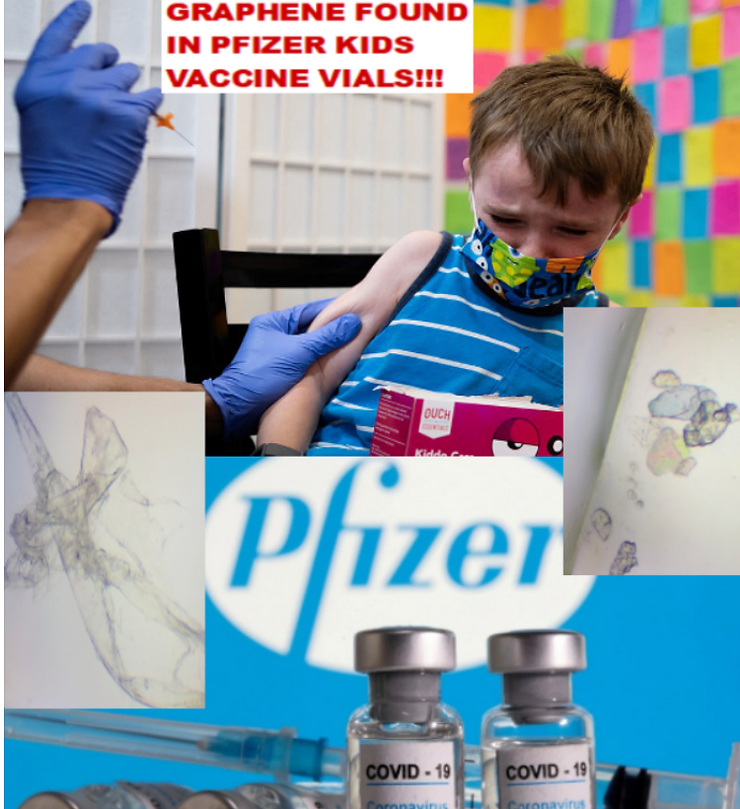
If you have been inoculated with the Pfizer VAXXXINE you may have been injected with a biochip of reduced graphene attached with ferric oxide and the eggs of the Trypanosoma Cruzi parasite! Check out the micrographs below!


Graphene Ferric oxide crystals (Ferromagnetic Properties) and Trypansoma cruzi parasite eggs were observed in the live capillary blood from a VAXXinated male using Brightfield, pHase contrast microscopy and confirmed with UV absorbance and Fluorescence Spectroscopy, Scanning Electron Microscopy, Transmission Electron Microscopy, Energy Dispersive Spectroscopy, X-ray Diffractometer and Nuclear Magnetic Resonance instruments. – Copyright Hikari Omni Media – Robert O. Young MSc, DSc, PhD, Naturopathic Practitioner – 2021

Ferromagnetic Properties of Graphene based Ferric oxide for Connection of the Human Body and Mind up to the Internet of Medical Things and the Internet of Things

Here is to PDF File for the Entire Article:

Graphene oxide is considered toxic (cytotoxic) in the smallest amounts and accumulates in the body and yet it is used everywhere, including the lipid-coated nanotubes that deliver vaccines and other medicines. It is also mutagenic, causing DNA damage and continuing mutation, just as mRNA vaccines have recently been proven to do.
Graphene Oxide as nanowebs can combine dots, flakes, tubes, and sheets into animated nanowebs that self-organize, self-replicate and direct the building of tissue-like material in the circulatory system, as well as nanocircuits that target certain organs (brain, heart, ovaries, testes, liver, etc.) and carry the payload inside the nanotube to the targeted organ. All of these things are already happening, these are not science predictions, they are science fact. These types of inhuman, mechanical, anti-life systems are being used right now, approved by the FDA, CDC, NIH, WHO, AMA, etc., to target and attack cancer cells in a variety of organs.
Doctors can inject and move large amounts of Graphene Oxide with a magnet to a specific organ, inject a hydrogel and control the release of more nano-tubes from the hydrogel with a phone app to conduct telemedicine. These GO nan-technologies, merged with mRNA, create the most-deadly genocide in human history because we have only seen the initial results. Some estimates are that over twelve million have died directly associated with the current jab. Untold others have terrible adverse reactions. That doesn’t take into consideration the millions who have died due to the same vaccine death shots given out continuously for flu, pneumonia, shingles, or childhood vaccines.
The vaXXXine induced Sudden Infant Death Syndrome is now joined by the Sudden Adult Death Syndrome and the medical authorities actively look the other way as hundreds of athletes drop dead on the playing field before the audience. And still, the vaccine has not been pulled from the market and the guilty prosecuted.
As red blood cells are poisoned and destroyed by graphene oxide, luciferase, PEG, Parasites and genetically modified RNADNA genetic/microzymian fragments from mouse, bat, monkey and aborted fetuses contained in the CoVid-19 so-called vaccines all blood counts begin to dangerously drop!

This includes dangerously low red blood cell counts, hemoglobin, hematocrit, white blood cell counts, including neutrophils, basophils, eosinophils, T and B lymphocytes all begin to drop below normal ranges because people are being poisoned by each toxic injection.
The more injections one receives the more destruction to the blood leading to VAIDS or Vaccine Acquired Immunodeficiency Syndrome leading to organ, gland and connective tissue degeneration and death.

Graphene Oxide as fullerenes (buckyballs) is, as yet, little used. It is a man-made 3D structure folded from GO sheets which can also make other 3D geometric solids. This is totally different than C-60, a natural occurring substance which is presumed to be from meteorites. Naturally occurring C-60 (fullerenes/buckyballs), which can be found in the rare mineraloid shungite, has nothing to do with the GO buckyballs created in laboratories.
Scientists believe that bucky-balls (C-60) originate in space and are a highly developed form of carbon exposed to cosmic heat. There is also C-70, C-80, and other carbon compounds found in space that have exposed carbon to tremendous heat, which is one way to create Graphene Oxide – burn a steak on your barbeque and you have simple graphene oxide.
It seems quite likely that as human intelligence develops, so too does carbon develop in its many organic forms through a natural process of metamorphoses. We owe our life to carbon and if more perfect forms of carbon already exist in our solar system and cosmos, then obviously we can metamorphose carbon into higher forms and functions.
Unfortunately, our voodoo witch-doctor mad-scientists haven’t considered any of these ideas as they are actively devolving into a “Graphene World” of one, two, and three dimensional, man-made monstrosities by injected a Frankenstein mutation (mRNA is mutagenic) into the new graphene oxide, genetically modified human being, a new species that has fallen out of the 3D world into the 2D graphene World.
Humans can advance to an objective view of time and enter a world of spiritual endurance (4D) instead of the illusion of linear time (3D). Modern materialistic science has devolved into two dimensional nanowebs that mimic human neural nets with 2D nanosheets/nanowebs that build “fake” human tissue with their 1D nano graphene dust/graphene flakes that are designed to kill human beings, ultimately leading to 0D – death. This is clearly planned elimination of everyone who does not know the secret –
“Don’t take any injection of any kind.”
The Graphene World is a world of sub-nature, a step backwards into immoral animal, plant, and mineral realms, not a step forward into higher forms of carbon in super-nature that are part of human ascension.
Graphene Oxide is a man-made sub-element that can only lead into darkness and the horrifying medical genocide we are seeing around us in all fields of medicine. Every person involved in gain of function research on deadly manmade synthetic viruses and so-called vaXXXines is an enemy to humanity.
Using GO to deliver any vaXXXine is diabolical, then add mRNA and Trypanosoma cruzi parasites and you have a truly evil group of murderers.
These types of experiments on “uninformed” humans are creating a new species of ill and dying humanity and an elite Harmaceutical syndicate that openly advocates depopulation by injection, toxic food, toxic chem-trail air, a medical industry creating illness, economic slavery, psychological subliminal programming, and the mass hypnosis of media propaganda that sold the world a fake plandemic – the fear of manmade Virus XXX.
Manmade Virus XXX, the highly prophesied pandemic of huge proportions, is spliced into the synthetic manmade virus that was created in a bioweapon P-4 lab and disseminated to all other P-4 secure biolabs throughout the world.
This biological weapon of mass destruction was bio-engineered with funding from Dr. Anthony Fauci and the National Institutes of Health, the CDC, and the World Health Organization of the United Nation. The United Nations is a clear Anti-American war-actor that took over OUR American Constitutional freedoms via Big Harmaceutical Terrorism with a fake plandemic supported with lies and sick protocols that has already killed tens of millions of men, women, boys, girls, children, babies and unborn babies in the womb. It was all made possible by a corrupt Congress passing laws that allowed it: The All Hazards and Plandemic Act of 2019.
Ferric oxide as a vaccine adjuvant has been in most childhood vaXXXines since 2008 absent of ANY ‘safe and effective’ scientific research.
Graphene oxide is present everywhere in the environment and yet is poisonous, as proven in every study of its toxicity.
Toxicity of Graphene Family Nanomaterials

Toxicity of Graphene Family of Nanomaterials

Toxicity of Graphene Family of Nanomaterials in Cell Models

Toxicity of Graphene of Family of Nanomaterials in Cell Models

And yet the medical industry pushes forward without any moral reflection on the harm being done to humans. These Big Harma doctors and drug-pushers are individuals who have devolved into immoral animals who are now below even what an animal would do to another animal.
The demonic forces involved in this global Harmaceutical World War III are quite real and wish to turn all humans into machine-augmented cyborgs who can be “stopped” or “controlled” by pushing a button that activates transhuman networks inside the human body.
This nefarious plan has been patented by Richard C. Walker and is called “The Aggressive Remote Control of Everything”, which can only be fully accomplished by having an “OFF” button on every human being created by nano graphene technology!
What is Graphene Oxide (GO)?

Graphene Oxide (GO) is a single atom carbon layer where both surfaces of the layer are modified by oxygen containing functional groups that are bonded together in a repeating pattern of hexagons. There is tremendous interest in graphene and its derivatives [graphene oxide (GO) and reduced GO (rGO)] due to their superior mechanical, thermal, electrical, optical, and chemical-adsorption properties. In the past few years, graphene-based materials attracted much attention and were used for many practical applications in various industries. Recent developments on graphene synthesis from foodstuffs, use of graphene for food analyses, and graphene-based analytical methods in detection (e.g., composition, contaminants, toxins, and volatile organic compounds) are used to help to ascertain the quality and/or safety of foods. There are also antibacterial properties of graphene-based nanomaterials and their applications in food packaging.
Graphene Family Nano-materials trigger local and systemic toxic effects, induce genotoxicity in vitro and in vivo, alter the gut microbiome, cause genetic mutations, and are inedible. Further toxicological and risk assessment studies are needed especially when used in food or injections of any type.
Different applications have been suggested for graphene nano-materials (GFNs) in the food and feed chain. However, it is necessary to perform a risk assessment before they become market-ready, and when consumer exposure is demonstrated. For this purpose, the European Food Safety Authority has published a guidance that has been recently updated to identify and characterize toxicological hazards related to GFNs after oral exposure. GFNs seemed to resist gastrointestinal digestion and were not able to be absorbed, distributed, and excreted, inducing toxic effects at different levels, including genotoxicity. Also, dose has an important role as it has been reported that low doses are more toxic than high doses because GFNs tend to aggregate in the digestive system, changing the internal exposure scenario. Thus, further studies including a thorough toxicological evaluation are required to protect humanity from the, as yet unknown, effects of GFNs.
Although Graphene Oxide – like graphene – is also a 2 Dimensional material, its properties are very different from that of graphene. It does not absorb visible light, has a lower electric conductance compared to that of graphene, and demonstrates significantly higher chemical activity. Its high electron mobility is 100x faster than silicon; it conducts heat 2x better than diamond; its electrical conductivity is 13x better than copper; it absorbs only 2.3% of reflecting light; it is impervious so that even the smallest atom can’t pass through a defect-free monolayer graphene sheet with a thickness of about 0.33 nanometers. There are about 3 million layers of graphene in a 1 mm thick sheet of graphite. Harder than diamond yet more elastic than rubber; tougher than steel yet lighter than aluminum – graphene is the strongest known material.

Some of the most promising applications of graphene are publicized as being in electronics (as transistors and interconnects), detectors (as sensor elements) and thermal management. The first graphene field-effect transistors (FETs) have already been created and used for nano analog communication or nano digital applications.
An ever-increasing number of research groups are exploiting programmable self-assembly properties of nucleic acids in creating rationally designed nano-shapes, nano-machines, and nano-electronic devices that can self-assemble for many different uses. These devices include nano-routers, nano-antennas, and nano-circuit boards. Medical nano-technology researchers have created nano-bots, a popular term for molecules with a unique property that enables them to be programmed to carry out a specific task.
When Graphene Oxide is injected into the body and interacts with biological blood or tissue, the GO picks up hydrogen and becomes graphene hydroxide.
The OH (hydroxy) groups can then split off a proton which leaves a negative charge affecting the whole graphene sheet and making it highly acidic and damaging to red blood cells. It also is incredibly sharp and acts like razor blades cutting blood vessels, tissue, and organs. Self-organizing GO tubes and sheets can block capillaries and arteries, with devastating effects when this occurs in the heart and lungs.
Graphene Oxide inside the body causes thrombogenicity, blood clotting, post inflammatory syndrome or systemic or multi-organ inflammations, causes alteration of the immune system, collapse of the immune system, cytokine storms, neurodegeneration, and mutagenic effects changing the DNA of the host.


Inhaled Graphene Oxide spreads evenly throughout the alveolar tract and causes bilateral pneumonias, inflammation of the mucous membranes, and loss of taste and smell.
Graphene Oxide toxicity in the human body behaves like SARS-CoV-2, generating the same symptomatology.
Graphene, Graphene Oxide (GO), carbon nano-tubes, and the entire graphene-family nano-materials (GFN) are toxic in almost all their forms, causing mutagenesis (cancer, chromosomal alteration), cell death, apoptosis, necrosis, and the release of free radicals.

It creates immunosuppression, damage to the central nervous system, circulatory, endocrine, reproductive, and urinary systems, which can cause anaphylactic death, and multi-organ dysfunction. It increases toxicity rapidly in the lungs, creating cytokine storms leading to bilateral pneumonia, genotoxicity, and DNA damage.
Several typical mechanisms underlying Graphene Oxide nano-material’s toxicity have been revealed in numerous studies including my own, for instance, physical destruction, oxidative stress, DNA damage, inflammatory response, apoptosis, autophagy, and necrosis. In these mechanisms, toll-like receptors, transforming growth factor-beta (TGF-β) and tumor necrosis factor-alpha (TNF-α) dependent-pathways are involved in the signaling pathway network, and oxidative stress plays a crucial role in these pathways.

Many experiments have shown that Graphene Oxide nano-materials have toxic side effects in many biological applications.
According to the USA FDA, graphene, Graphene Oxide, and reduced graphene oxide elicit toxic effects both in vitro and in vivo.
Graphene-family nano-materials (GFN) are not approved by the USA FDA for human consumption.
The inventor of graphene oxide says the vaccine will kill you – Dr. Mylo Canderian, Ph.D. [born Milos Iskanderianos, Corfu, Greece, 1938], who developed the patent for Graphene Oxide for use as a Hematological Bioweapon in 2015.

Graphene Oxide has been used in a wide variety of nano-medical applications including tissue engineering, cancer treatment, medical imaging, and drug delivery.
Its physiochemical properties allow for a structure to regulate the behavior of stem cells, with the potential to assist in the intracellular delivery of DNA, growth factors, and synthetic proteins. Due to its unique behavior in biological environments, GO is used in cancer therapies. It has also been used in vaccines and immunotherapy, including as a dual-use adjuvant and carrier of biomedical nano materials, including graphene oxide.
In September 2020, researchers at the Shanghai National Engineering Research Center for Nanotechnology in China filed a patent for use of Graphene Oxide in a recombinant vaccine under development against SARS-CoV-2.

The properties of graphene are exceptional from a physical, thermodynamic, electronic, mechanical, and magnetic point of view. Its characteristics allow it to be used as a superconductor, crystallized graphene nano-antenna, and graphene quantum dot nano-routers as seen in the blood of the vaXXXinated.

Graphene Oxide Nano Antennas and Routers

It is an electromagnetic wave absorbing material, a signal emitter-receiver, and an antenna which makes it possible to create advanced nano and micrometric scale electronics.
Graphene is a radio-Modula table nano-material. The graphene molecule also has the ability to inject electrons into other biological substances depending on the electromagnetic environment and temperature. Graphene is activated at room temperature and above.
Graphene can multiply radiation, acting as a nano-antenna, or else a signal repeater, a transistor. Exposure to electromagnetic radiation can cause the exfoliation of the material into smaller particles called Graphene Quantum Dots (GQD), whose properties and physical peculiarities are enhanced since they act by amplifying electromagnetic signals and, with that, the emission distance, especially in environments such as the human body.
Graphene quantum dots as seen below can acquire various morphologies like hexagonal, triangular, circular, bucky-bulls, or irregular polygons and geometric solids.

The nightmare of Graphene Oxide circuits in human food is a Frankenstein monster that kills
As Mark Wilson’s headline reads: “Graphene Is Here, And Electronics In Your Food Are Coming.” Mark’s article highlights the research conducted by Jeff Rice University that uses a stock laser to carve edible circuits into food. These researchers have successfully used a commercial laser to transform the surface carbon in foods – like toast, coconuts shells, potatoes, and Girl Scout cookies – into graphene.
Without using any special vacuums or clean rooms, graphene can be patterned into an impossibly thin, edible circuit.
Graphene can be used to help fuel cells to store power, radio hardware to transmit data, glowing elements to light up, and all sorts of sensors, as well as deliver a preprogrammed piece of toast that can control your body.
These graphene circuits resemble a dark, inky tattoo, a bit like very burnt toast. But, don’t forget, graphene is inedible, toxic, and a nerve poison.
Graphene Ferric Oxide Technology

Iron oxide nano-structures (IONs) in combination with graphene or its derivatives – e.g., Graphene Oxide and reduced graphene oxide – hold great promise toward engineering of efficient nano-composites for enhancing the performance of advanced devices in many applicative fields.
Due to the peculiar electrical and electrocatalytic properties displayed by composite structures in nanoscale dimensions, increasing efforts have been directed in recent years toward tailoring the properties of IONs-graphene based nanocomposites for developing more efficient electrochemical sensors.
Unique features of IONs e.g., strong magnetic properties, low toxicity, high adsorption ability for immobilization of desired biomolecules and good biocompatibility, together with elegant properties of this new member of the carbon family e.g., high electrical/thermal conductivity, large surface area and electrocatalytic properties, have stimulated many interests for overcoming difficulties in realizing new scientific ideas or improving the performance of many current devices and methods.

Catalytic activity of the graphene-IONs can be improved due to enhanced electronic communication e.g., charge transfer between catalyst and support. Additionally, synergistic effects of graphene sheets and IONs components provide nano-composite with novel physicochemical properties and consequently enhance electrochemical performance. As a result, graphene-IONs nano-composites have been considered as one of the most promising hybrid materials that can boost the development of more efficient electrochemical sensors.
Hydrogels and Graphene Oxide
Due to their tissue-like mechanical properties, hydrogels are being increasingly used for biomedical applications; a well-known example are soft contact lenses. These gel-like polymers consist of 90 percent water, are elastic and particularly biocompatible.
Hydrogels that are also electrically conductive allow additional fields of application, for example in the transmission of electrical signals in the body or as sensors.
Graphene and graphene derivatives (e.g., Graphene Oxide (GO) reduced graphene oxide (rGO)) have been incorporated into hydrogels to improve the properties (e.g., mechanical strength) of conventional hydrogels and/or develop new functions (e.g., electrical conductivity and drug loading/delivery). Unique molecular interactions between graphene derivatives and various small or macromolecules enable the fabrication of various functional hydrogels appropriate for different biomedical applications.
In order to produce electrically conductive hydrogels, conventional hydrogels are usually mixed with current-conducting nano-materials that are made of metals or carbon, such as gold nano-wires, graphene or carbon nano-tubes.

PEER-REVIEWED PUBLISHED RESEARCH, STUDIES & INTERVIEWS
To demonstrate the truth and efficacy of the above statements concerning the graphene family materials, we present below a series of research projects which summarize the “state of the art” concerning research in Graphene Oxide in its many forms. Much of what has been said above may have sounded alarmist, or even like wild, sci-fi fairytales of transhumanism, but the research below demonstrates that all of the experiments on humans with graphene substances has been going on for many years on a massive scale.
The “innovations” in nano-particle research are not “illegal” but should certainly be “not allowed” by any moral scientist, doctor, or sane person.
For the sake of innovation, humanity is now a collective lab rat to be experimented on by morally bankrupt drug-doctors preaching the Gospel of Transhuman manipulation of the building blocks of DNA, human organs, tissue creation, neurological control through wetworks, and inhuman mechanical thinking that dominates “precision medicine” and nano-biology.
Essentially, nano-biology should be an oxymoron instead of the current medical, experimental treatment, vaccine, or deadly medical procedure. Man-made toxic graphene does not belong in the human body.
After reading these studies and watching a few interviews, I believe you will agree that all Graphene Oxide use must end immediately and parties guilty of these heinous crimes against humanity must be brought to justice.
Graphene and Ferric Oxide in Vaccines
From Published Research: Hikari Omni Media Publications, February 5th, 2021, “Scanning & Transmission Electron Microscopy Reveals Graphene & Parasites in CoV-19 Vaccines”, R.O. Young.

Abstract: Currently there are four major pharmaceutical companies who manufacture a SARS-CoV-2 now called SARS-CoV-19 vaccine. These manufactures and their vaccine are Pfizer–BioNTech mRNA Vaccine, the Moderna-Lonza mRNA-1273 Vaccine, the Serum Institute Oxford Astrazeneca Vaccine and the Janssen COVID -19 Vaccine, manufactured by Janssen Biotech Inc., a Janssen Pharmaceutical Company of Johnson & Johnson, a recombinant, replication-incompetent adenovirus type 26 expressing the SARS-CoV-2 spike protein.

The intended purpose of these vaccines are to provide immunity from the so-called infectious novel coronavirus or SARS-CoV – 2 virus now called the SARS-CoV – 19. These four pharmaceutical companies have not provided complete FDA disclosure on their vaccine box, insert fact sheet or label for many of the major and/or minor ingredients contained within these so-called vaccines. The purpose of this research article is to identify those specific major and minor ingredients contained in the Pfizer VaXXXine, the Moderna VaXXXine, the Astrazeneca VaXXXine and the Janssen VaXXXine using various scientific anatomical, physiological and functional testing for each SARS-COV-2-19 vaccine.

As a human right, governed under World Law by the Nuremberg Code of 1947, the vaccine specific ingredient information is critical, required and necessary to know so that any human from any country in the World can make an informed decision whether or not to consent to the SAR-CoV-2-10-19 inoculation. We have conducted the scientific testing on each vaccine and have identified several ingredients or adjuvants that have not been disclosed which are contained in these four SARS-CoV – 2 -19 vaccines.
Currently, these vaccines are being administered to millions of humans around the World under an Emergency Use Authorization (EUA) issued by each country without full disclosure of all ingredients and in some cases mandated by governments or employers in violation of individual human rights under the Nuremberg Code of 1947.


Graphene and Ferric Oxide in VaXXXines
From Published Reserach: ACS Publications, February 17, 2021, “In Situ Transforming RNA Nanovaccines from Polyethylenimine Functionalized Graphene Oxide Hydrogel for Durable Cancer Immunotherapy”, Yue Yin, Xiaoyang Li, Haixia Ma, Jie Zhang, Di Yu, Ruifang Zhao, Shengji Yu, Guangjun Nie, and Hai Wang

Abstract: Messenger RNA (mRNA) vaXXXine is a promising candidate in cancer immunotherapy as it can encode tumor-associated antigens with an excellent safety profile.
Unfortunately, the inherent instability of RNA and translational efficiency are major limitations of the mRNA vaccines. Here, we report an injectable hydrogel formed with graphene oxide (GO) and polyethylenimine, which can generate mRNA and adjuvants (R848)-laden nanovaXXXines for at least 30 days after subcutaneous injection.
The released nanographene vaXXXines can protect the mRNA from degradation and confer targeted delivering capacity to lymph nodes. The data shows that this transformable hydrogel can significantly increase the number of antigen-specific CD8+ T cells theoretically inhibiting the tumor growth with only one treatment is a scientific illusion and fraud.
Meanwhile, this nanographene hydrogel theoretically generates an antigen specific antibody in the serum which in turn prevents the occurrence of metastasis which is the illusion of its authors. Collectively, these pseudo results demonstrate a theoretical potential of the PEI-functionalized GO transformable hydrogel for effective cancer immunotherapy. This theory is totally fallacious since cancer is an acidic disease of the interstitial fluids of the Interstitium and NOT a disease of the cells themselves!

The Food and Drug Administration (FDA) has unfortunately approved many types of iron oxide nanoparticles for clinical use, such as treating iron deficiency, contrast agents for magnetic resonance imaging (MRI) and drug delivery platforms.
In one study, researchers explored the combined use of iron oxide nanoparticles (superparamagnetic Fe3O4 nanoparticles) as a vaXXXine delivery platform and immune potentiator, and investigated how this formulation affected cytokine expression in macrophages and dendritic cells (DCs) in vitro and tumor growth in vivo. Their highly toxic iron oxide nanoparticles greatly promoted the activation of immune cells and cytokine production because the body fluids were poisoned, inducing potent humoral and cellular immune responses simply due to the systemic poisoning with graphene ferric oxide.

These results suggest that this nanoparticle-based delivery system has strong potential to cause harm by polluting the interstitial fluids of the Interstitium and should never be utilized as a vaXXXine for any cancerous condition or for a manmade gain of function virus which will only injury and eventually kill the host!
Superparamagnetic iron oxide nanoparticles (SPIONs) as a contrast agent have been widely used in magnetic resonance imaging for tumor diagnosis and theragnostic and is cytotoxic, genotoxic and magnetic toxic to the glands, organs and tissues of the human and animal body.
This is why there has been serious safety concerns of SPIONs with cirrhosis of the liver related to excess iron-induced oxidative stress or systemic poisoning of the interstitial and vascular fluids of the blood and Interstitium organ.
Analysis with PCR array of the toxicity pathways revealed the high dose of SPIONs induced significant expression changes of a distinct subset of genes in the cirrhosis liver.
All these results suggested that excess iron of the high dose of SPIONs is a risk factor for liver cirrhosis because of the marked impacts of elevated lipid metabolism, disruption of iron homeostasis and possibly, aggravated loss of liver functions.
At present, nanoparticles are being used for various biomedical applications where they facilitate laboratory diagnostics and therapeutics. More specifically for drug delivery purposes, the use of nanoparticles is attracting increasing attention due to their unique capabilities and their negligible side effects not only in cancer therapy but also in the treatment of other ailments. Among all types of nanoparticles, biocompatible superparamagnetic iron oxide nanoparticles (SPIONs) with proper surface architecture and conjugated targeting ligands/proteins have attracted a great deal of attention for drug delivery applications.
Superparamagnetic iron oxide nanoparticles (SPIONs) have drawn attention because of their excellent superparamagnetic properties such as controllable size, large surface area-to-volume ratio, and nontoxicity. Surface functionalization of SPIONs with therapeutic molecules, including antimicrobial agents, has been successfully used in nanomedicine.
Through application of an external magnetic field, antimicrobial-loaded SPIONs can be guided to the desired outfectious site allowing a direct and specific questionable and concerning so-called therapeutic effect. The great advantage of SPIONs is their magnetic properties that allow direct delivery of matter into the targeted zone without testing the toxic effects to the interstitial fluids potentially causing more harm then good..
When infused intravenously, these SPIONs can be used to detect and characterize small focal lesions in the liver. They also can be administered orally in order to visualize the digestive tract, and can be used as biomarkers to evaluate the efficacy of treatments. But still further investigations are required using labeled SPIONs in the field of molecular imaging since they are a direct assault on the alkaline integrity of the body ocean of interstitial fluids that surrounds every cell in the human body.
Superparamagnetic iron oxide nanoparticles (SPIONs) have been studied for various biomedical applications, such as contrast agents, iron replacement therapies, drug delivery, tissue repair, hyperthermia, cell and tissue targeting, and transfection. SPIONs have an iron oxide core that is coated by an organic or inorganic layer. Bare SPIONs may be toxic because there is chemical reactive, so the coating layer prevents aggregation and agglomeration of the nanoparticles and reduces iron oxide oxidation. SPIONs are largely studied for magnetic resonance imaging and targeted delivery of drug and antigen to the required sites.
SPIONs have been approved by the FDA for treatment of anemia in adult patients with chronic renal disease. SPIONs are also used for noninvasive diagnosis of chronic liver diseases, nonalcoholic steatohepatitis, cirrhosis, liver tumors, magnetic resonance angiography, lymph node imaging, bone marrow imaging, and atherosclerotic plaque imaging.
Iron oxide Nanoparticles in Food
From Published Research: Science of Food, November 20, 2017, “Is nano safe in foods? Establishing the factors impacting the gastrointestinal fate and toxicity of organic and inorganic food-grade nanoparticles”, David Julian McClements & Hang Xiao

Nanotechnology offering the food industry a number of new approaches for improving the quality, shelf life, safety, and healthiness of foods. Nevertheless, there is concern from consumers, regulatory agencies, and the food industry about potential adverse effects (toxicity) associated with the application of nanotechnology in foods.
In particular, there is concern about the direct incorporation of engineered nanoparticles into foods, such as those used as delivery systems for colors, flavors, preservatives, nutrients, and nutraceuticals, or those used to modify the optical, rheological, or flow properties of foods or food packaging. This review article summarizes the application of both inorganic (silver, iron oxide, titanium dioxide, silicon dioxide, and zinc oxide) and organic (lipid, protein, and carbohydrate) nanoparticles in foods, highlights the most important nanoparticle characteristics that influence their behavior, discusses the importance of food matrix and gastrointestinal tract effects on nanoparticle properties, emphasizes potential toxicity mechanisms of different food-grade nanoparticles, and stresses important areas where research is still needed. The authors note that nanoparticles are already present in many natural and processed foods, and that new kinds of nanoparticles may be utilized as functional ingredients by the food industry in the future.
Nanotechnology can be utilized to improve food quality, shelf life, safety, cost, and nutritional benefits. In some cases, the nanomaterials used in the food industry are not intended to find their way into the final food product, e.g., those used in packaging, sensors, and antimicrobial treatments designed for sanitizing food manufacturing plants.
Engineered nanoscale materials (ENMs) may be intentionally added to foods or they may inadvertently find their way into foods (such as nanoparticles in packaging materials that leach into the food matrix). ENMs may be used to create delivery systems for nutrients, nutraceuticals, colors, flavors, and preservatives, or they may be used to modify the texture, appearance, or stability of foods. Nanoscale structures may be present in foods as the result of routinely used food processing operations, such as homogenization, grinding, and cooking.
Nanoparticles present in foods can be categorized as either organic or inorganic. Inorganic materials, such as silver, iron oxide, titanium dioxide, silicon dioxide, or zinc oxide are commonly used. These particles are either crystalline or amorphous solids at ambient temperature, which may be spherical or non-spherical, have different surface characteristics and coatings, and come in different sizes depending on the initial materials and preparation conditions used in their fabrication.
Inorganic nanoparticles:
Silver nanoparticles are used as antimicrobial agents in foods and food packaging materials.
Zinc oxide nanoparticles may be used as a source of zinc and in food packaging as antimicrobial agents to prevent contamination of foods and as ultraviolet light absorbers.
Iron oxide nanoparticles are utilized in foods as colorants or sources of bioavailable iron and come in different sizes, shapes, and crystalline forms.
Titanium dioxide nanoparticles are used as functional ingredients in certain foods to provide characteristic optical properties such as increased lightness and brightness
.
Silicon dioxide nanoparticles are added to certain powdered foods as anticaking agents to enhance flow properties, e.g., salts, icing sugar, spices, dried milk, and dry mixes.
Organic nanoparticles
Lipid nanoparticles are widely present within many commercial food products, like beverage emulsions, such as soft drinks, fortified waters, fruit juices, and dairy drinks, contain small oil droplets dispersed in water.
Protein nanoparticles are the casein micelles found in bovine milk and other dairy products, which are small clusters of casein molecules and calcium phosphate ions.
Carbohydrate nanoparticles are typically assembled from digestible or indigestible polysaccharides, such as starch, cellulose, alginate, carrageenan, pectin, and xanthan and they may be indigestible within the upper gastrointestinal tract (GIT).
Some organic substances used to fabricate food nanoparticles (such as dietary fibers and mineral oils) may not be digested in the upper GIT. Inorganic nanoparticles are also not digested in the GIT. Any nanoparticles that are not digested or absorbed in the upper GIT will reach the lower GIT where they may alter the microbiome in a negative way. The ability of inorganic nanoparticles to produce toxicity is often associated with their chemical reactivity, which depends on their composition. For example, some inorganic nanoparticles dissolve and release ions that promote undesirable chemical or biochemical reactions (e.g., silver nanoparticles).
Ingested nanoparticles accumulate in numerous tissues!
These nanoparticles travel across the mucus layer and are then absorbed by active or passive transport mechanisms.
After they have been absorbed into the cells, they accumulate within the cells. The accumulation of nanoparticles within specific tissues may lead to long-term problems if they exhibit toxic effects above a certain accumulation threshold. This mechanism of action is likely to be most important for inorganic nanoparticles that are bio-persistent (not normally digested or metabolized in GIT).
Nanoparticles may produce toxicity in cells through a variety of different mechanisms
One of the most important factors contributing to the toxicity of inorganic nanoparticles is their ability to generate reactive oxygen species (ROS), such as singlet oxygen, superoxide, hydrogen peroxide and hydroxyl radicals. These ROS may then cause damage to cell membranes, organelles, and the nucleus by interacting with lipids, proteins, or nucleic acids. As a result, many biochemical functions required to maintain cell viability, such as ATP production, DNA replication, and gene expression, may be adversely affected. A number of studies have reported the ability of inorganic nanoparticles to increase the generation of ROS in cells and to produce cytotoxicity.
The ability of nanoparticles to greatly increase the oral bioavailability of hydrophobic substances does have adverse health effects by promoting the uptake of undesirable non-polar substances in foods, such as certain pesticides (glyphosates, etc.) and hormones. For example, a food product that contains lipid nanoparticles (such as a beverage, sauce, dressing, or cream) may increase the bioavailability of hydrophobic pesticides on fruits or vegetables consumed with them.
Graphene Self-Assembles into Blood Vascular Structures
From Published Research: Materials Today Connecting the Materials Community, March 19, 2020, “New graphene-based material self-assembles into vascular structures.”

An international team of scientists, led by Alvaro Mata at the University of Nottingham and Queen Mary University London in the UK, has discovered a new material that can be 3D printed to create tissue-like vascular structures. In a paper in Nature Communications, the scientists report developing a way to 3D print graphene oxide with a protein that can organize into tubular structures that replicate some of the properties of vascular tissue.
“This work offers opportunities in bio-fabrication by enabling simultaneous top-down 3D bioprinting and bottom-up self-assembly of synthetic and biological components in an orderly manner from the nanoscale,” said Mata. “Here, we are bio-fabricating micro-scale capillary-like fluidic structures that are compatible with cells, exhibit physiologically relevant properties, and have the capacity to withstand flow.
This could enable the recreation of vasculature in the lab and have implications in the development of safer and more efficient drugs, meaning treatments could potentially reach patients much more quickly.”
Self-assembly is the process by which multiple components spontaneously organize into larger, well-defined structures
Biological systems rely on this process to controllably assemble molecular building blocks into complex and functional materials exhibiting remarkable properties such as the capacity to grow, replicate and perform robust functions. The new biomaterial is produced by the self-assembly of a protein with graphene oxide. This self-assembly process allows the flexible regions of the protein to order and conform to the graphene oxide, generating a strong interaction between them. By controlling the way in which the two components are mixed, it is possible to guide their assembly at multiple scales in the presence of cells to produce complex robust structures.
Self-Assembling Graphene Nanotubes
From Published Research: Angewandte Chemie, First published: March 14, 2001, “Self-Assembling Organic Nanotubes,” T. Bong,Thomas D. Clark Dr., Juan R. Granja Prof. Dr., M. Reza Ghadiri Prof.


Hollow tubular structures of molecular dimensions perform diverse biological functions in nature. Examples include scaffolding and packaging roles played by cytoskeletal microtubules and viral coat proteins, respectively, as well as the chemical transport and screening activities of membrane channels. In the preparation of such tubular assemblies, biological systems make extensive use of self-assembling and self-organizing strategies. Owing to numerous potential applications in areas such as chemistry, biology, and materials science considerable effort has recently been devoted to preparation of artificial nanotubular structures.
This article reviews design principles and the preparation of synthetic organic nanotubes, with special emphasis on noncovalent processes such as self-assembly and self-organization.
Programmable Living Systems Based on a Foundation of Graphene Oxide
From Published Research: Nature Reviews Materials, “Materials design by synthetic biology,” Tzu-Chieh Tang, Bolin An, Yuanyuan Huang, Sangita Vasikaran, Xiaoyu Jiang

Synthetic biology applies genetic tools to engineer living cells and organisms analogous to the programming of machines. In materials synthetic biology, engineering principles from synthetic biology and materials science are integrated to redesign living systems as dynamic and responsive materials with emerging and programmable functionalities.
In this Review, we discuss synthetic-biology tools, including genetic circuits, model organisms and design parameters, which can be applied for the construction of smart living materials. We investigate non-living and living self-organizing multifunctional materials, such as intracellular structures and engineered biofilms, and examine the design and applications of hybrid living materials, including living sensors, therapeutics and electronics, as well as energy-conversion materials and living building materials. Finally, we consider prospects and challenges of programmable living materials and identify potential future applications.
Engineered Living Materials
From Published Research: MIT Libraries, “Towards engineering living functional materials,” 2021, Tang, Tzu-Chieh,Ph. D.Massachusetts Institute of Technology

The field of engineered living materials (ELMs) aims to recapitulate the remarkable properties of natural biology to create novel, growable, multifunctional materials using genetically engineered organisms. Most relevant pioneering work was created using nano- to microscale biofilm [GO], which has rather small yields and usually requires costly modification.
Second, releasing genetically modified microorganisms (GMMs) into the field for food, water, or agricultural applications is often considered risky due to the uncertainty of wild-type organisms acquiring undesirable traits, such as antibiotic resistance, from the GMMs. A significant effort in addressing these unmet needs is called for. This Thesis starts with an introduction of genetic circuits and an in-depth review of the current trends in materials synthetic biology, which includes two major categories of ELMs: self-organizing functional materials and hybrid living materials. The following chapters describe the technologies developed to achieve high scalability and safe deployment of ELMs in these two categories and living devices suitable for real-world applications.
Graphene Oxide Toxicity
From Published Research: Biomedical Research International, Volume 2021 |Article ID 5518999, “Synthesis and Toxicity of Graphene Oxide Nanoparticles: A Literature Review of In Vitro and In Vivo Studies”, Asmaa Rhazouani, Halima Gamrani , Mounir El Achaby , Khalid Aziz, Lhoucine Gebrati, Md Sahab Uddin, and Faissal AZIZ, https://doi.org/10.1155/2021/5518999

Nanomaterials have been widely used in many fields in the last decades, including electronics, biomedicine, cosmetics, food processing, buildings, and aeronautics. The application of these nanomaterials in the medical field could improve diagnosis, treatment, and prevention techniques. Graphene oxide (GO), an oxidized derivative of graphene, is currently used in biotechnology and medicine for cancer treatment, drug delivery, and cellular imaging. Also, GO is characterized by various physicochemical properties, including nanoscale size, high surface area, and electrical charge.
However, the toxic effect of GO on living cells and organs is a limiting factor that limits its use in the medical field.
Recently, numerous studies have evaluated the biocompatibility and toxicity of GO in vivo and in vitro. In general, the severity of this nanomaterial’s toxic effects varies according to the administration route, the dose to be administered, the method of GO synthesis, and its physicochemical properties.
Graphene nanoparticles are widely used in electronics, aeronautics, energy, agriculture, cosmetics, medicine, textile production, and many other fields. They are currently used to administer drugs, proteins, genes, vaccines, polypeptides, and nucleic acids. GO is a nanomaterial that has been known for more than 150 years and is used in many applications. In recent years, graphene has been exploited in the medical field, particularly for DNA sequencing, the development of biosensors, and cell differentiation and growth.
As graphene is insoluble in water, its applications are limited to passive platforms for detection and cell work. Its functional derivative GO has unique properties that make it more effective for biomedical applications. It is characterized by its ability to disperse in many solvents, facilitating its handling.
In addition, GO is used to administer anticancer drugs in biological cells, aptamers for ATP probing in epithelial cells, and gene delivery. These nanomaterials have a large surface area and can maintain drugs’ stability without altering the biological activity, much more than other nanomaterials.
GO is characterized by properties that make it attractive in other areas such as sensors and energy storage. As applications increase, exposure to GO increases across populations. These include exposures during nanomaterial manufacturing and biomedical treatment. GO is involved in many applications, but there is one main factor limiting “its toxicity” limiting its use. Researchers are often faced with the problem of balancing the positive therapeutic effects of GO with the side effects associated with its toxicity
Graphene Oxide as a Vaccine Carrier and Adjuvant
From Published Research: Acta Biomaterialia, Volume 112, August 2020, Pages 14-28, “Recent progress of graphene oxide as a potential vaccine carrier and adjuvant,” WanjunCaoab, LinHea, Weidong Caob, Xiaobing HuangaKun, Jiac Jingying Dai
Adjuvants and carriers have been appropriately added to the vaccine formulation to improve the immunogenicity (effective chemical poisoning) of the antigen and induce long-lasting immunity which is a theory of scientism!
Graphene oxide (GO), widely employed for the delivery of biomolecules, excels in loading and delivering antigen and shows the potentiality of activating the immune system.
However, GO aggregates in biological liquid [blood clots] and induces cell death, and it also exhibits poor bio-solubility and bio-compatibility.
To address these limitations, various surface modification protocols have been employed to integrate aqueous compatible substances with GO to effectively improve its biocompatibility. More importantly, these modifications render functionalized-GO with superior properties as both carriers and adjuvants.
Due to its unique physicochemical properties, graphene oxide is widely employed in medicine for purposes of photothermal treatment of cancer, drug delivery, antibacterial therapy, and medical imaging. This reserach describes the surface modification of graphene oxide and for the first time summarizes that functionalized graphene oxide serves as a vaccine carrier and shows significant adjuvant activity in activating cellular and humoral immunity.
Precision medicine informs us that graphene oxide has been studied for its promising uses in a wide variety of nanomedical applications including tissue engineering, cancer treatment, medical imaging, and drug delivery.
Its physiochemical properties allow for a structure to regulate the behavior of stem cells, with the potential to assist in the intracellular delivery of DNA, growth factors, and synthetic proteins that could allow for the repair and regeneration of muscle tissue.
Due to its unique behavior in biological environments, graphene oxide has also been proposed as a novel material in early cancer diagnosis.
It has also been explored for its uses in vaccines and immunotherapy, including as a dual-use adjuvant and carrier of biomedical materials.
Several typical mechanisms underlying graphene oxide nanomaterial’s toxicity have been revealed, for instance, physical destruction, oxidative stress, DNA damage, inflammatory response, apoptosis, autophagy, and necrosis. In these mechanisms, toll-like receptors (TLR), transforming growth factor-beta (TGF-β) and tumor necrosis factor-alpha (TNF-α) dependent-pathways are involved in the signaling pathway network, and oxidative stress plays a crucial role in these pathways.
Many experiments have shown that graphene oxide nanomaterials have toxic side effects in many biological applications. According to the USA FDA, graphene, graphene oxide, and reduced graphene oxide elicit toxic effects both in vitro and in vivo.
Graphene-family nanomaterials (GFN) are not approved by the USA FDA for human consumption.
Graphene Oxide-incorporated Hydrogels in Medicine
From Published Research: Polymer Journal, Volume 52, pages 823-837, May 8, 2020, “Graphene oxide-incorporated hydrogels for biomedical applications,” Jongdarm Yi, Goeun Choe, Junggeon Park Young Lee.

Graphene and graphene derivatives (e.g., graphene oxide) have been incorporated into hydrogels to improve the properties (e.g., mechanical strength) of conventional hydrogels and/or develop new functions (e.g., electrical conductivity and drug loading/delivery). Unique molecular interactions between graphene derivatives and various small or macromolecules enable the fabrication of various functional hydrogels appropriate for different biomedical applications. In this mini-review, we highlight the recent progress in GO-incorporated hydrogels for biomedical applications while focusing on their specific uses as mechanically strong materials, electrically conductive scaffolds/electrodes, and high-performance drug delivery vehicles.
Graphene Oxide Used in So-Called Pfizer, Moderna, Astrazenica and J&J Gain of Function So-Called Vaccines
From Published Research: “Nanoscale, Functionalized graphene oxide serves as a novel vaccine nano-adjuvant for robust stimulation of cellular immunity”, Ligeng Xu, Jian Xiang, Ye Liu, Jun Xu, Yinchan Luo, Liangzhu Feng, Zhuang Liu and Rui Peng
Benefiting from their unique physicochemical properties, graphene derivatives have attracted great attention in biomedicine. In this study, we carefully engineered graphene oxide (GO) as a vaccine adjuvant for immunotherapy using urease B (Ure B) as the model antigen. Our work not only presents a novel, highly effective GO-based vaccine nano-adjuvant, but also highlights the critical roles of surface chemistry for the rational design of nano-adjuvants.
STILL NOT CONVINCED THAT GRAPHENE OXIDE AND FERRIC OXIDE ARE IN THE VAXXXINES? THEN CHECK OUT THE FOLLOWING PEERED-REVIEWED JOURNAL ARTICLES
NEED MORE PROOF!
Article 1
August 2021
Graphene oxide (GO) nanomaterials have significant advantages for drug delivery and electrode materials in neural science, however, their exposure risks to the central nervous system (CNS) and toxicity concerns are also increased. The current studies of GO-induced neurotoxicity remain still ambiguous, let alone the mechanism of how complicated GO chemistry affects its biological behavior with neural cells. In this study, we characterized the commercially available GO in detail and investigated its biological adverse effects using cultured SH-SY5Y cells. We found that ultrasonic processing in medium changed the oxidation status and surface reactivity on the planar surface of GO due to its hydration activity, causing lipid peroxidation and cell membrane damage. Subsequently, ROS-disrupted mitochondrial homeostasis, resulting from the activation of NOX2 signaling, was observed following GO internalization. The autophagy-lysosomal network was initiated as a defensive reaction to obliterate oxidative damaged mitochondria and foreign nanomaterials, which was ineffective due to reduced lysosomal degradation capacity. These sequential cellular responses exacerbated mitochondrial stress, leading to apoptotic cell death. These data highlight the importance of the structure-related activity of GO on its biological properties and provide an in-depth understanding of how GO-derived cellular redox signaling induces mitochondrion-related cascades that modulate cell functionality and survival.
Article 2
Graphene and its derivatives (for example, nanoscale graphene oxide (NGO)) have emerged as extremely attractive nanomaterials for a wide range of applications, including diagnostics and therapeutics. In this work, we present a systematic study on the in vivo distribution and pulmonary toxicity of NGO for up to 3 months after exposure. Radioisotope tracing and morphological observation demonstrated that intratracheally instilled NGO was mainly retained in the lung. NGO could result in acute lung injury (ALI) and chronic pulmonary fibrosis. Such NGO-induced ALI was related to oxidative stress and could effectively be relieved with dexamethasone treatment. In addition, we found that the biodistribution of 125I-NGO varied greatly from that of 125I ions, hence it is possible that nanoparticulates could deliver radioactive isotopes deep into the lung, which might settle in numerous ‘hot spots’ that could result in mutations and cancers, raising environmental concerns about the large-scale production of graphene oxide.
Article 3
April 2017
We summarized the findings of toxicity studies on graphene-based nanomaterials (GNMs) in laboratory mammals. The inhalation of graphene (GP) and graphene oxide (GO) induced only minimal pulmonary toxicity. Bolus airway exposure to GP and GO caused acute and subacute pulmonary inflammation. Large-sized GO (L-GO) was more toxic than small-sized GO (S-GO). Intratracheally administered GP passed through the air-blood barrier into the blood and intravenous GO distributed mainly in the lungs, liver, and spleen. S-GO and L-GO mainly accumulated in the liver and lungs, respectively. Limited information showed the potential behavioral, reproductive, and developmental toxicity and genotoxicity of GNMs. There are indications that oxidative stress and inflammation may be involved in the toxicity of GNMs. The surface reactivity, size, and dispersion status of GNMs play an important role in the induction of toxicity and biodistribution of GNMs. Although this review paper provides initial information on the potential toxicity of GNMs, data are still very limited, especially when taking into account the many different types of GNMs and their potential modifications. To fill the data gap, further studies should be performed using laboratory mammals exposed using the route and dose anticipated for human exposure scenarios.
Article 4
May 2014
Graphene nanoparticle dispersions show immense potential as multifunctional agents for in vivo biomedical applications. Herein, we follow regulatory guidelines for pharmaceuticals that recommend safety pharmacology assessment at least 10-100 times higher than the projected therapeutic dose, and present comprehensive single dose response, expanded acute toxicology, toxicokinetics, and respiratory/cardiovascular safety pharmacology results for intravenously administered dextran-coated graphene oxide nanoplatelet (GNP-Dex) formulations to rats at doses between 1 and 500 mg/kg. Our results indicate that the maximum tolerable dose (MTD) of GNP-Dex is between 50 mg/kg ≤ MTD < 125 mg/kg, blood half-life < 30 min, and majority of nanoparticles excreted within 24 h through feces. Histopathology changes were noted at ≥250 mg/kg in the heart, liver, lung, spleen, and kidney; we found no changes in the brain and no GNP-Dex related effects in the cardiovascular parameters or hematological factors (blood, lipid, and metabolic panels) at doses < 125 mg/kg. The results open avenues for pivotal preclinical single and repeat dose safety studies following good laboratory practices (GLP) as required by regulatory agencies for investigational new drug (IND) application
Article 5
July 2021
Nanomaterials have been widely used in many fields in the last decades, including electronics, biomedicine, cosmetics, food processing, buildings, and aeronautics. The application of these nanomaterials in the medical field could improve diagnosis, treatment, and prevention techniques. Graphene oxide (GO), an oxidized derivative of graphene, is currently used in biotechnology and medicine for cancer treatment, drug delivery, and cellular imaging. Also, GO is characterized by various physicochemical properties, including nanoscale size, high surface area, and electrical charge. However, the toxic effect of GO on living cells and organs is a limiting factor that limits its use in the medical field. Recently, numerous studies have evaluated the biocompatibility and toxicity of GO in vivo and in vitro. In general, the severity of this nanomaterial’s toxic effects varies according to the administration route, the dose to be administered, the method of GO synthesis, and its physicochemical properties. This review brings together studies on the method of synthesis and structure of GO, characterization techniques, and physicochemical properties. Also, we rely on the toxicity of GO in cellular models and biological systems. Moreover, we mention the general mechanism of its toxicity.
Article 6
July 2020
Being a member of the nanofamily, carbon nanomaterials exhibit specific properties that mostly arise from their small size. They have proved to be very promising for application in the technical and biomedical field. A wide spectrum of use implies the inevitable presence of carbon nanomaterials in the environment, thus potentially endangering their whole nature. Although scientists worldwide have conducted research investigating the impact of these materials, it is evident that there are still significant gaps concerning the knowledge of their mechanisms, as well as the prolonged and chronic exposure and effects. This manuscript summarizes the most prominent representatives of carbon nanomaterial groups, giving a brief review of their general physico-chemical properties, the most common use, and toxicity profiles. Toxicity was presented through genotoxicity and the activation of the cell signaling pathways, both including in vitro and in vivo models, mechanisms, and the consequential outcomes. Moreover, the acute toxicity of fullerenol, as one of the most commonly investigated members, was briefly presented in the final part of this review. Thinking small can greatly help us improve our lives, but also obliges us to deeply and comprehensively investigate all the possible consequences that could arise from our pure-hearted scientific ambitions and work.
Article 7
March 2016
Recently, graphene and graphene-related materials have attracted a great deal of attention due their unique physical, chemical, and biocompatibility properties and to their applications in biotechnology and medicine. However, the reports on the potential toxicity of graphene oxide (GO) in biological systems are very few. The present study investigated the response of kidneys in male Sprague-Dawley rats following exposure to 0, 10, 20 and 40 mg/Kg GO for five days. The results showed that administration of GOs significantly increased the activities of superoxide dismutase, catalase and glutathione peroxidase in a dose-dependent manner in the kidneys compared with control group. Serum creatinine and blood urea nitrogen levels were also significantly increased in rats intoxicated with GO compared with the control group. There was a significant elevation in the levels of hydrogen peroxide and lipid hydro peroxide in GOs-treated rats compared to control animals. Histopathological evaluation showed significant morphological alterations of kidneys in GO-treated rats compared to controls. Taken together, the results of this study demonstrate that GO is nephrotoxic and its toxicity may be mediated through oxidative stress. In the present work, however, we only provided preliminary information on toxicity of GO in rats; further experimental verification and mechanistic elucidation are required before GO widely used for biomedical applications.
Article 8
October 2016
Carbon-based functional nanomaterials have attracted immense scientific interest from many disciplines and, due to their extraordinary properties, have offered tremendous potential in a diverse range of applications. Among the different carbon nanomaterials, graphene is one of the newest and is considered the most important. Graphene, a monolayer material composed of sp2-hybridized carbon atoms hexagonally arranged in a two-dimensional structure, can be easily functionalized by chemical modification. Functionalized graphene and its derivatives have been used in diverse nano-biotechnological applications, such as in environmental engineering, biomedicine, and biotechnology. However, the prospective use of graphene-related materials in a biological context requires a detailed comprehension of these materials, which is essential for expanding their biomedical applications in the future. In recent years, the number of biological studies involving graphene-related nanomaterials has rapidly increased. These studies have documented the effects of the biological interactions between graphene-related materials and different organizational levels of living systems, ranging from biomolecules to animals. In the present review, we will summarize the recent progress in understanding mainly the interactions between graphene and cells. The impact of graphene on intracellular components, and especially the uptake and transport of graphene by cells, will be discussed in detail.
Article 9
July 2016
Despite graphene being proposed for a multitude of biomedical applications, there is a dearth in the fundamental cellular and molecular level understanding of how few-layer graphene (FLG) interacts with human primary cells. Herein, using human primary umbilical vein endothelial cells as model of vascular transport, we investigated the basic mechanism underlying the biological behavior of graphene. Mechanistic toxicity studies using a battery of cell based assays revealed an organized oxidative stress paradigm involving cytosolic reactive oxygen stress, mitochondrial superoxide generation, lipid peroxidation, glutathione oxidation, mitochondrial membrane depolarization, enhanced calcium efflux, all leading to cell death by apoptosis/necrosis. We further investigated the effect of graphene interactions using cDNA microarray analysis and identified potential adverse effects by down regulating key genes involved in DNA damage response and repair mechanisms. Single cell gel electrophoresis assay/Comet assay confirmed the DNA damaging potential of graphene towards human primary cells.
Article 10
December 2016
Despite the rapid expansion of the biomedical applications of graphene oxide (GO), safety issues related to GO, particularly with regard to its effects on vascular endothelial cells (ECs), have been poorly evaluated. To explore possible GO-mediated vasculature cytotoxicity and determine lateral GO size relevance, we constructed four types of GO: micrometer-sized GO (MGO; 1089.9 ± 135.3 nm), submicrometer-sized GO (SGO; 390.2 ± 51.4 nm), nanometer-sized GO (NGO; 65.5 ± 16.3 nm), and graphene quantum dots (GQDs). All types but GQD showed a significant decrease in cellular viability in a dose-dependent manner. Notably, SGO or NGO, but not MGO, potently induced apoptosis while causing no detectable necrosis. Subsequently, SGO or NGO markedly induced autophagy through a process dependent on the c-Jun N-terminal kinase (JNK)-mediated phosphorylation of B-cell lymphoma 2 (Bcl-2), leading to the dissociation of Beclin-1 from the Beclin-1–Bcl-2 complex. Autophagy suppression attenuated the SGO- or NGO-induced apoptotic cell death of ECs, suggesting that SGO- or NGO-induced cytotoxicity is associated with autophagy. Moreover, SGO or NGO significantly induced increased intracellular calcium ion (Ca2+) levels. Intracellular Ca2+chelation with BAPTA-AM significantly attenuated microtubule-associated protein 1A/1B-light chain 3-II accumulation and JNK phosphorylation, resulting in reduced autophagy. Furthermore, we found that SGO or NGO induced Ca2+ release from the endoplasmic reticulum through the PLC β3/IP3/IP3R signaling axis. These results elucidate the mechanism underlying the size-dependent cytotoxicity of GOs in the vasculature and may facilitate the development of a safer biomedical application of GOs.
Article 11
January 2018
The successful applications of graphene nanomaterials in nanobiotechnology and medicine as well as their effective translation into real clinical utility hinge significantly on a thorough understanding of their nanotoxicological profile. Of all aspects of biocompatibility, the hemocompatibility of graphene nanomaterials with different blood constituents in the circulatory system is one of the most important elements that needs to be well elucidated. Once administered into biological systems, graphene nanomaterials may inevitably come into contact with the surrounding plasma proteins and blood cells. Crucially, the interactions between these hematological entities and graphene nanomaterials will influence the overall efficacy of their biomedical applications. As such, a comprehensive understanding of the hemotoxicity of graphene nanomaterials is critically important. This review presents an up-to-date elucidation of the hemotoxicity of graphene nanomaterials through their interactions with blood proteins and cells, as well as offers some perspectives on the current challenges, opportunities, and future development of this important field.
Article 12
January 2014
The raised considerable concerns about the possible environmental health and safety impacts of graphene nanomaterials and their derivatives originated from their potential widespread applications. We performed a comprehensive study about biological interaction of grapheme nanomaterials, specifically in regard to its differential surface functionalization (oxidation status), by using OMICS in graphene oxide (GO) and reduced graphene oxide (rGO) treated HepG2 cells. Differential surface chemistry (particularly, oxidation – O/C ratio) modulates hydrophobicity/philicity of GO/rGO which in turn governs their biological interaction potentiality. Similar toxic responses (cytotoxicity, DNA damage, oxidative stress) with differential dose dependency were observed for both GO and rGO but they exhibited distinct mechanism, such as, hydrophilic GO showed cellular uptake, NADPH oxidase dependent ROS formation, high deregulation of antioxidant/DNA repair/apoptosis related genes, conversely, hydrophobic rGO was found to mostly adsorbed at cell surface without internalization, ROS generation by physical interaction, poor gene regulation etc. Global gene expression and pathway analysis displayed that TGFβ1 mediated signaling played the central role in GO induced biological/toxicological effect whereas rGO might elicited host-pathogen (viral) interaction and innate immune response through TLR4–NFkB pathway. In brief, the distinct biological and molecular mechanisms of GO/rGO were attributed to their differential surface oxidation status.
Article 13
2015
Graphene oxide (GO) is a promising nanomaterial for application in a variety of biomedical fields, including neuro-oncology, neuroimaging, neuroregeneration and drug delivery. Microglia are the central macrophage-like cells critically involved in neuroimmunity. However, the interaction between GO and microglia remained mostly unknown. The present study investigated the influence of GO on the production of proinflammatory cytokines by microglia. Primary murine microglial cells were treated with GO (1–25 μg/mL) followed by stimulation with lipopolysaccharide (LPS) for 24 h. The cell viability was measured by spectrophotometry using AlamarBlueⓇ. The levels of interleukin (IL)-1β and tumor necrosis factor (TNF)-α in the supernatants were measured by enzyme-linked immunosorbent assay (ELISA). The IL-1β converting enzyme (ICE) activity was measured using a specific fluorescent substrate. The activity of cathepsin B and the lysosomal permeability and alkalinity were determined by flow cytometry. Treatment with GO did not affect cell viability, but significantly suppressed the production of IL-1β. In contrast, the production of TNF-α was unaltered. In addition, the lysosomal permeability and alkalinity in microglia treated with GO were increased, whereas the activity of cathepsin B and ICE was decreased. Collectively, these results demonstrated that exposure to GO differentially affected the production of proinflammatory cytokines, which is associated with the modulation of the lysosomal pathway of cytokines processing.
Article 14
January 2017
Increased production volumes and a broadening application spectrum of graphene have raised concerns about its potential adverse effects on human health. Numerous reports demonstrate that graphene irrespective of its particular form exerts its effects on a widest range of living organisms, including prokaryotic bacteria and viruses, plants, micro- and macro-invertebrates, mammalian and human cells and whole animals in vivo. However, the available experimental data is frequently a matter of significant divergence and even controversy. Therefore, we provide here a critical analysis of the most recent (2015–2016) reports accumulated in the graphene-related materials biocompatibility and toxicology field in order to elucidate the cutting edge achievements, emerging trends and future opportunities in the area. Experimental findings from the diverse in vitro and in vivo model systems are analysed in the context of the most likely graphene exposure scenarios, such as respiratory inhalation, ingestion route, parenteral administration and topical exposure through the skin. Key factors influencing the toxicity of graphene and its complex derivatives as well as potential risk mitigation approaches exploiting graphene physicochemical properties, surface modifications and possible degradation pathways are also discussed along with its emerging applications for healthcare, diagnostics and innovative therapeutic approaches.
Article 15
January 2014
Graphene and its derivatives are promising candidates for important biomedical applications because of their versatility. The prospective use of graphene-based materials in a biological context requires a detailed comprehension of the toxicity of these materials. Moreover, due to the expanding applications of nanotechnology, human and environmental exposures to graphene-based nanomaterials are likely to increase in the future. Because of the potential risk factors associated with the manufacture and use of graphene-related materials, the number of nanotoxicological studies of these compounds has been increasing rapidly in the past decade. These studies have researched the effects of the nanostructural/biological interactions on different organizational levels of the living system, from biomolecules to animals. This review discusses recent results based on in vitro and in vivo cytotoxicity and genotoxicity studies of graphene-related materials and critically examines the methodologies employed to evaluate their toxicities. The environmental impact from the manipulation and application of graphene materials is also reported and discussed. Finally, this review presents mechanistic aspects of graphene toxicity in biological systems. More detailed studies aiming to investigate the toxicity of graphene-based materials and to properly associate the biological phenomenon with their chemical, structural, and morphological variations that result from several synthetic and processing possibilities are needed. Knowledge about graphene-based materials could ensure the safe application of this versatile material. Consequently, the focus of this review is to provide a source of inspiration for new nanotoxicological approaches for graphene-based materials.
Article 16
April 2018
With the vigorous development of nanometer-sized materials, nanoproducts are becoming widely used in all aspects of life. In medicine, nanoparticles (NPs) can be used as nanoscopic drug carriers and for nanoimaging technologies. Thus, substantial attention has been paid to the potential risks of NPs. Previous studies have shown that numerous types of NPs are able to pass certain biological barriers and exert toxic effects on crucial organs, such as the brain, liver, and kidney. Only recently, attention has been directed toward the reproductive toxicity of nanomaterials. NPs can pass through the blood–testis barrier, placental barrier, and epithelial barrier, which protect reproductive tissues, and then accumulate in reproductive organs. NP accumulation damages organs (testis, epididymis, ovary, and uterus) by destroying Sertoli cells, Leydig cells, and germ cells, causing reproductive organ dysfunction that adversely affects sperm quality, quantity, morphology, and motility or reduces the number of mature oocytes and disrupts primary and secondary follicular development. In addition, NPs can disrupt the levels of secreted hormones, causing changes in sexual behavior. However, the current review primarily examines toxicological phenomena. The molecular mechanisms involved in NP toxicity to the reproductive system are not fully understood, but possible mechanisms include oxidative stress, apoptosis, inflammation, and genotoxicity. Previous studies have shown that NPs can increase inflammation, oxidative stress, and apoptosis and induce ROS, causing damage at the molecular and genetic levels which results in cytotoxicity. This review provides an understanding of the applications and toxicological effects of NPs on the reproductive system.
Article 17
2014
Concentration-dependent cyto and genotoxicities of graphene oxide (GO) and reduced GO (rGO) sheets on spermatozoa were studied. rGO sheets with various surface chemical states were achieved using hydrazine (N2H4) hydrothermal (HT) reactions and green tea polyphenols (GTPs). Although 0.1 μg mL−1 graphene could not change sperm viability and kinetic parameters, <40% and 20% of spermatozoa were viable and progressively motile, after 2 h incubation with 400 μg mL−1 GO or rGO, respectively. All the graphene nanomaterials induced concentration-dependent reductions of adenosine triphosphate and NAD+/NADH produced by spermatozoa for motility and metabolic activity. While GO, N2H4–rGO, and HT-rGO sheets caused increasing reactive oxygen species and sperm nitric oxide production, GO sheets reduced by antioxidant GTPs decreased them. Hence, physical trapping of spermatozoa by graphene (particularly GTP–rGO) is one of the important mechanisms describing the cytotoxicity, in addition to the other reactions, resulting in the inactivation and/or death of spermatozoa. Graphene genotoxicity was initiated by 1.0 μg mL−1 of N2H4–rGO and HT-rGO and 10 μg mL−1 of GO and GTP–rGO sheets. The extremely sharp edge and/or high mobility of N2H4–rGO provided easy penetration of the sheets into spermatozoa to interact with cell nuclei. In contrast, the steric effect induced by GTPs attached on rGO caused a lower genotoxicity.
Article 18
Short-term in vivo exposure to graphene oxide can cause damage to the gut and testis – ScienceDirect
April 2017
Graphene oxide (GO) has unique physicochemical properties and also has a potentially widespread use in every field of daily life (industry, science, medicine). Demand for nanotechnology is growing every year, and therefore many aspects of its toxicity and biocompatibility still require further clarification.
This research assesses the in vivo toxicity of pure and manganese ion-contaminated GO that were administrated to Acheta domesticus with food (at 200 mg kg−1 of food) throughout their ten-day adult life.
Our results showed that short-term exposure to graphene oxide in food causes an increase in the parameters of oxidative stress of the tested insects (catalase – CAT, total antioxidant capacity – TAC), induces damage to the DNA at a level of approximately 35% and contributes to a disturbance in the stages of the cell cycle and causes an increase of apoptosis. Moreover, upon analyzing histological specimens, we found numerous degenerative changes in the cells of the gut and testis of Acheta domesticus as early as ten days after applying GO.
A more complete picture of the GO risk can help to define its future applications and methods for working with the material, which may help us to avoid any adverse effects and damage to the animal.
Article 19
December 2015
In vivo dose-dependent effects of nanoscale graphene oxide (NGO) sheets on reproduction capability of Balb/C mice were investigated. Biodistribution study of the NGO sheets (intravenously injected into male mice at dose of ∼2000 μg/mL or 4 mg/kg of body weight) showed a high graphene uptake in testis. Hence, in vivo effects of the NGO sheets on important characteristics of spermatozoa (including their viability, morphology, kinetics, DNA damage and chromosomal aberration) were evaluated. Significant in vivo effects was found at the injected concentrations ≥200 μg/mL after (e.g., ∼45% reduction in sperm viability and motility at 2000 μg/mL). Observation of remarkable DNA fragmentations and chromosomal aberrations of the spermatozoa after ∼8 weeks from the first weekly injection were assigned to the involvement of the NGO in spermatogenesis of the mice. The uptake of the NGO in the testis could also increase the generation of reactive oxygen species in semen of the mice. Moreover, semen of the NGO-treated mice (containing the damaged spermatozoa) might disturb the hormone secretion and pregnant functionality of female mice (∼44, 35 and 59% reduction in fertility, gestation ability and multi-production capability) and also viability of the next generation (∼15% reduction in postnatal viability of delivered pups).
Article 20
August 2016
Carbon-based nanomaterials such as single-walled carbon nanotubes and reduced graphene oxide are currently being evaluated for biomedical applications including in vivo drug delivery and tumor imaging. Several reports have studied the toxicity of carbon nanomaterials, but their effects on human male reproduction have not been fully examined. Additionally, it is not clear whether the nanomaterial exposure has any effect on sperm sorting procedures used in clinical settings. Here, we show that the presence of functionalized single walled carbon nanotubes (SWCNT-COOH) and reduced graphene oxide at concentrations of 1–25 μg/mL do not affect sperm viability. However, SWCNT-COOH generate significant reactive superoxide species at a higher concentration (25 μg/mL), while reduced graphene oxide does not initiate reactive species in human sperm. Further, we demonstrate that exposure to these nanomaterials does not hinder the sperm sorting process, and microfluidic sorting systems can select the sperm that show low oxidative stress post-exposure.
Article 21
July 2019
Graphene, a two-dimensional carbon sheet with single-atom thickness, shows immense promise in several nanoscientific and nanotechnological applications, including in sensors, catalysis, and biomedicine. Although several studies have shown the cytotoxicity of graphene oxide in different cell types, there are no comprehensive studies on human embryonic kidney (HEK293) cells that include transcriptomic analysis and an in vitro investigation into the mechanisms of cytotoxicity following exposure to graphene oxide. Therefore, we exposed HEK293 cells to different concentrations of graphene oxide for 24 h and performed several cellular assays. Cell viability and proliferation assays revealed a significant dose-dependent cytotoxic effect on HEK293 cells. Cytotoxicity assays showed increased lactate dehydrogenase (LDH) leakage and reactive oxygen species (ROS) generation, and decreased levels of reduced glutathione (GSH) and increased level of oxidized glutathione indicative of oxidative stress. This detailed mechanistic approach showed that graphene oxide exposure elicits significant decreases in mitochondrial membrane potential and ATP synthesis, as well as in DNA damage and caspase 3 activity. Furthermore, our RNA-Seq analysis revealed that HEK293 cells exposed to graphene oxide significantly altered the expression of genes involved in multiple apoptosis-related biological pathways. Moreover, graphene oxide exposure perturbed the expression of key transcription factors, promoting these apoptosis-related pathways by regulating their downstream genes. Our analysis provides mechanistic insights into how exposure to graphene oxide induces changes in cellular responses and massive cell death in HEK293 cells. To our knowledge, this is the first study describing a combination of cellular responses and transcriptome in HEK293 cells exposed to graphene oxide nanoparticles, providing a foundation for understanding the molecular mechanisms of graphene oxide-induced cytotoxicity and for the development of new therapeutic strategies.
Article 22
May 2010
Graphitic nanomaterials such as graphene layers (G) and single-wall carbon nanotubes (SWCNT) are potential candidates in a large number of biomedical applications. However, little is known about the effects of these nanomaterials on biological systems. Here we show that the shape of these materials is directly related to their induced cellular toxicity. Both G and SWCNT induce cytotoxic effects, and these effects are concentration- and shape-dependent. Interestingly, at low concentrations, G induced stronger metabolic activity than SWCNT, a trend that reversed at higher concentrations. Lactate dehydrogenase levels were found to be significantly higher for SWCNT as compared to the G samples. Moreover, reactive oxygen species were generated in a concentration- and time-dependent manner after exposure to G, indicating an oxidative stress mechanism. Furthermore, time-dependent caspase 3 activation after exposure to G (10 μg/mL) shows evidence of apoptosis. Altogether these studies suggest different biological activities of the graphitic nanomaterials, with the shape playing a primary role.
Article 23
February 2021
Arguments regarding the biocompatibility of graphene-based materials (GBMs) have never ceased. Particularly, the genotoxicity (e.g., DNA damage) of GBMs has been considered the greatest risk to healthy cells. Detailed genotoxicity studies of GBMs are necessary and essential. Herein, we present our recent studies on the genotoxicity of most widely used GBMs such as graphene oxide (GO) and the chemically reduced graphene oxide (RGO) toward human retinal pigment epithelium (RPE) cells. The genotoxicity of GO and RGOs against ARPE-19 (a typical RPE cell line) cells was investigated using the alkaline comet assay, the expression level of phosphorylated p53 determined via Western blots, and the release level of reactive oxygen species (ROS). Our results suggested that both GO and RGOs induced ROS-dependent DNA damage. However, the DNA damage was enhanced following the reduction of the saturated C–O bonds in GO, suggesting that surface oxygen-containing groups played essential roles in the reduced genotoxicity of graphene.
Article 24
October 2016
Polyethylene glycol (PEG) coating has been frequently used to improve the pharmacokinetic behavior of nanoparticles. Studies that contribute to better unravel the effects of PEGylation on the toxicity of nanoparticle formulation are therefore highly relevant. In the present study, reduced graphene oxide (rGO) was functionalized with PEG, and its effects on key components of the blood–brain barrier, such as astrocytes and endothelial cells, were analyzed in culture and in an in vivo rat model. The in vitro studies demonstrated concentration-dependent toxicity. The highest concentration (100 μg/mL) of non-PEGylated rGO had a lower toxic influence on cell viability in primary cultures of astrocytes and rat brain endothelial cells, while PEGylated rGO induced deleterious effects and cell death. We assessed hippocampal BBB integrity in vivo by evaluating astrocyte activation and the expression of the endothelial tight and adherens junctions proteins. From 1 h to 7 days post-rGO-PEG systemic injection, a notable and progressive down-regulation of protein markers of astrocytes (GFAP, connexin-43), the endothelial tight (occludin), and adherens (β-catenin) junctions and basal lamina (laminin) were observed. The formation of intracellular reactive oxygen species demonstrated by increases in the enzymatic antioxidant system in the PEGylated rGO samples was indicative of oxidative stress-mediated damage. Under the experimental conditions and design of the present study the PEGylation of rGO did not improve interaction with components of the blood–brain barrier. In contrast, the attachment of PEG to rGO induced deleterious effects in comparison with the effects caused by non-PEGylated rGO.
To read additional 1750 peer-reviewed scientific articles on CoV VaXXXine poisoning go to:
https://www.drrobertyoung.com/post/1750-covid-vaxxine-publications-and-case-reports-citing-adverse-effects-post-covid-vaxxination
Want to Learn More?
The Identification of Trypanosoma Parasites in the Pfizer VAXXine!
Have you wondered why Pfizer had to deep-freeze their mRNA so-called CoViid – 19 VAXXine?So the Trypanosoma cruzi parasites do not hatch before injection into humans. The vector for parasite infection is from the so-called coronavirus injection!
How Did I Discover Parasites in the CoVid – 19 Vaccines?
Using phase contrast, darkfield and bright field microscopy and finally directed energy spectroscopy I observed and identified in the Pfizer VAXXINE, graphene and ferric oxide and the Trypanosoma cruzi parasite eggs.

A 0.5ml aqueous fraction image from Pfizer vaccine sample viewed under pHase contrast microscopy at 1000x magnification, showing a symplast of graphene oxide (upper left) next to a Trypanosoma cruzi parasite (lower right). Dr. Robert O. Young, Hikari Omni Publishing, September 11th, 2021.

A Scanning Electron Microscope Identifies the Blood-destroying, Blood-clotting Trypanosoma Cruzi Parasite in the Pfizer VaXXine and the Blood of the VAXXINATED!
Over 70 percent of the Western Hemisphere has been infected by the Transom blood sucking bug which will bite you like a mosquito and then defecate on your skin with Trypanosoma parasites and their eggs.

The over 70 percent infected in the Western Hemisphere has increased to over 90 percent infected because of the so-called coronavirus VAXXines and the intentional implantation of graphene, ferric oxide and Trypanosoma parasites!

If you are currently asymptomatic, there is a high risk with VAXXination and the implantation of magnetic ferric oxide attached to a base of reduced graphene creating a human quantum linked to 4G and 5G pulsating microwaves causing radiation poisoning and decompensated acidosis of the interstitial fluids of the Interstitium leading to pathological blood coagulation, oxygen deprivation, hypoxia and lung, heart, kidney, pancreas, spleen, liver, gallbladder, reproductive, thyroid, stomach, bowel and brain dis-ease.

The Interstitium Organ is the Largest Organ of the Human Body and its Interstitial Fluids Surround Every Body Cell! The Interstitium Organ Manages Cellular Health, Energy and Metabolic Waste. It is at the Core of Human Health and Fitness or Sickness and Disease! – https://www.drrobertyoung.com/blog/search/interstitial%20


The VAXXXination of the Coronavirus VAXXine containing Trypanosoma cruzi eggs directly into the interstitial fluids of the Interstitium and the eventual hatching of these eggs at body temperature, may lead to acute or chronic Chagas dis-ease caused by the very Trypansoma cruzi parasite identified and observed in the Pfizer coronavirus vaxxine.

Symptoms of Chagas or Trypanosoma Dis-Ease Include:
1. Fever 2. Headache 3. Body aches 4. Rash 5. Swelling around the eyelids
6. Enlarged heart 7. Heart failure 8. Stroke 9. Life-threatening ventricular arrhythmias 10. Cardiac arrest, and even more life threatening symptomologies listed below . . .

To learn more about the nondisclosed ingredients in the coronavirus vaxxines read my latest peer-reviewed published article by Acta Scientific Medical Science Journal, ASMS-22-RW-150 entitled “Scanning & Transmission Electron Microscopy Reveals Graphene Oxide in CoV-19 Vaccines” The PDF is attached below or you can click on the following link:
Watch, listen, learn and then SHARE with EVERYONE YOU LOVE & CARE ABOUT!

Special guest Dr. Robert Young makes his first appearance on the show to speak with Liberty Man about his microscopy and spectroscopy findings in the gene juice injections.

Dr. Young and Liberty Man talk about FDA corruption and share perspectives on the nanotechnology aspects of the shots, including topics such as RNA interference, toxicity, shedding, blood clots, and electromagnetic frequencies, and how all of this plays into the larger agenda of a digital ID social-credit prison system.
Support the work, research and findings of Dr. Robert O. Young at: https://www.givesendgo.com/research

No donation is too small and will be appreciated and used for continuous research, publications and public education!
* * *
As a special thank you for your donation, I will answer one health or lifestyle related question.
After your donation, please email me at: phmiraclelife@gmail.com
Keep your question short and very specific. Please include your first and last name, phone number and email. Your question will be answered within 72 hours.
God bless YOU ALL!
Stay healthy and happy! www.givesendgo.com/researchwww.drrobertyoung.com/blog